


Foamy microglia in MS brain
Researchers have discovered an important mechanism that may be linked to severe progression of multiple sclerosis (MS). In brain tissue from patients with rapidly progressing MS, they found large numbers of abnormal immune cells overloaded with fat droplets. The study offers new leads for treatments as well as biomarkers that could better predict disease progression.
In MS, the fatty insulating layer surrounding nerve fibers (myelin) is broken down in the brain and spinal cord. This can lead to neurological symptoms such as difficulties with walking and vision. MS progresses differently in every patient. Some people live for decades with relatively mild symptoms, while others become severely paralyzed at a young age. Researchers have therefore long tried to understand what causes these differences.
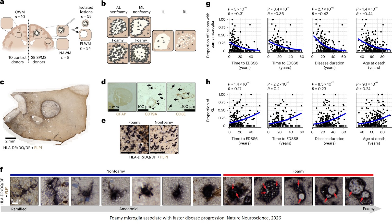
In the new study, the scientists focused on microglia: immune cells in the brain that clear waste and help repair damaged tissue. In MS patients, however, these cells change shape. They become filled with fat droplets, giving them a foamy appearance. Researchers call these cells “foamy microglia.” “We found that patients with large numbers of these foamy microglia had a more severe disease course more frequently,” says the researcher.
Under normal circumstances, microglia help clean up damage in the brain. In MS, however, this task may sometimes become too big. The researchers believe the cells absorb so much damaged myelin that they eventually become overwhelmed by their own waste-processing system. “These cells are probably trying to do something good: clearing up damage,” the author explains. “But they become overloaded, so to speak. As a result, they can no longer effectively contribute to repair.”
The foamy microglia/macrophages exhibit disrupted lipid metabolism, lysosomal stress and markers associated with heightened phagocytosis and antigen presentation without classical pro-inflammatory signatures.
These lesions are enriched for oxylipins, bismonoacylglycerolphosphates and cholesterol esters, and are associated with increased B cell infiltration and IgG1.
Monoacylglycerol lipase (MAGL), a lipid-metabolizing enzyme enriched in lesions with foamy microglia/macrophages, emerged as a potential therapeutic target. Inhibition of MAGL promoted lesion recovery and reduced microgliosis in a mouse model of demyelination. Alsp, oxylipins in cerebrospinal fluid correlate with the proportion of foamy lesions, suggesting potential biomarkers for progression.
The researchers also discovered that brain inflammations containing foamy microglia behave very differently at the molecular level from MS inflammations without these cells. For example, they contain specific fats involved in chronic inflammatory responses.
For a long time, inflammation was thought to be the driving force behind disease progression, but the study also shows that MS may be more complex than that alone. According to the researchers, their work points to a more subtle process. “It does not appear to be simply about the inflammatory response alone,” says the author. “These cells are probably attempting to clear damage and promote repair, but that process fails, worsens inflammation, and counteracts recovery.”
For the study, the scientists analysed brain tissue from 28 deceased MS patients who had donated their brains to the Netherlands Brain Bank. The team combined several advanced techniques that simultaneously examined gene activity, proteins, and fats within the same MS inflammatory lesions.
According to the researchers, the combination of modern technology and detailed knowledge of brain pathology was especially crucial. “Today we have incredibly sophisticated techniques that can map the brain in great detail,” the author says. “The technologies are fantastic, but they tell you relatively little if you cannot connect them to pathology in brain tissue. Precisely because brain tissue has been carefully studied and classified for years by the Netherlands Brain Bank, we were able to recognize these abnormal patterns.”
In the long term, the discovery may help improve predictions of MS disease progression. The researchers found indications that certain fats associated with foamy microglia can also be measured in patients’ cerebrospinal fluid. “That opens the possibility of developing biomarkers in the future that could help doctors identify earlier which patients are at risk of rapid decline — and which treatment would suit them best.”
In addition, the findings align with ongoing developments in new medicines targeting fat metabolism and chronic lesion expansion in MS. Some of these drugs are already being investigated in clinical studies in collaboration with Roche.
https://www.nature.com/articles/s41593-026-02302-3
https://sciencemission.com/Foamy-microglia