


Peptide ligase–mediated display
The technology for finding proteins and peptides that bind precisely to specific targets is a crucial foundation for developing new drugs and diagnosing diseases. To date, various methods called “display technologies” have been developed to search for proteins that bind to the desired target.
Recently, “cell-free protein synthesis,” a method that produces proteins in test tubes without using living cells like E. coli or yeast, has become widely used. This method allows for more flexible and efficient searches for target proteins since cell cultivation is unnecessary.
Many biological functions are regulated by the switching on and off of mechanisms triggered by the matching of a keyhole (receptor) formed by a protein's three-dimensional structure and a molecule (ligand) that fits perfectly into it. If this keyhole deforms (protein mutation) or if a false key is created, biological functions become disrupted, leading to disease.
Drug discovery research involves a process called screening to find compounds that fit into these receptor lock-and-key sites. The content published in this paper offers significant advantages, dramatically increasing the efficiency of this process and enabling the handling of highly toxic proteins that were previously difficult to work with.
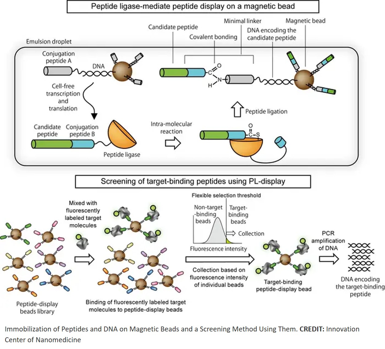
The underlying technology involves manufacturing and utilizing magnetic beads, each carrying a single peptide (a short-chain protein), without the need for cells. Traditional methods using living cells to produce peptides had drawbacks, such as inconsistent peptide quantities leading to unstable data. Compared to conventional approaches, this new method achieves over tenfold efficiency gains.
Furthermore, its ability to perform screening under high-temperature and high-salt conditions significantly reduces the time required for lead compound discovery in drug development. Details are provided below.
In this study, the authors utilized the properties of an enzyme called “peptide ligase,” which connects peptides together. Using this enzyme, they developed a new technique to firmly anchor peptides (short protein fragments) and their template DNA onto the surface of small beads via a short connecting segment consisting of just nine amino acids.
They named this method “PL-display.” The key feature of this technology is its ability to display one type of peptide and its corresponding DNA on a single bead without using any living cells. Furthermore, using an instrument called FACS, beads can be examined one by one, allowing precise selection based on numerical values of only those beads possessing the desired properties. To verify that this technology functions correctly, the authors conducted the following experiment.
First, the authors mixed equal amounts of DNA for two types of marker peptides, HA-tag and His-tag, and displayed each on beads. They
The results confirmed that only the genes for the targeted peptides were accurately extracted. Furthermore, even from a mixture containing only 0.01% (one in ten thousand) of the HA-tag gene, the authors succeeded in completely isolating the HA-tag gene with just one round of selection. Additionally, from approximately 1.7 million peptides with random sequences, they efficiently collected peptides binding to the anti-HA-tag antibody in just two rounds of selection.
By determining the strictness of recovery criteria based on the fluorescence intensity of each individual bead, they achieved the high efficiency of “10,000-fold concentration in a single sorting step,” which was difficult with conventional methods. This demonstrated that even from diverse peptide populations resembling those in actual drug discovery research, target peptides can be identified quickly.
This technology employs a method called “cell-free protein synthesis.” This is a method for producing proteins in test tubes without using living cells. Consequently, it is not constrained by the conditions required to grow cells and is unaffected by variations in how proteins are produced by individual cells.
Furthermore, it allows for the investigation of proteins that are harmful to cells or under special conditions different from the normal internal environment without issue. Additionally, in this technology, the protein and its template, DNA, are firmly connected by a strong bond that is difficult to break. Furthermore, each bead displays only one type of protein. This ensures stability even under harsh conditions like vigorous washing or high salt concentrations, allowing precise evaluation of “how strongly each bead binds to its target.”
Using FACS equipment, which sorts based on bead brightness (fluorescence intensity), allows researchers to freely and finely set criteria like “recovering only beads above a certain brightness threshold.”
This mechanism theoretically enables the selection not only of proteins with very strong binding, but also of proteins possessing “just the right binding strength”—neither too strong nor too weak.
Ultra-High-Throughput Screening of target-binding proteins using PL-display is expected to find applications across a wide range of fields, including drug discovery, diagnostics, and biomaterial development. These include the search for therapeutic and diagnostic peptides and low-molecular-weight antibodies, the development of industrial proteins for use in non-physiological environments, and high-speed screening through automation and robotics utilizing magnetic bead platforms.
https://academic.oup.com/pnasnexus/article/5/2/pgag031/8482226
https://sciencemission.com/Peptide-ligase%E2%80%93mediated-display