


Label-free and reporter-free minimally invasive Raman spectroscopy with deep simulation to detect brain cancer
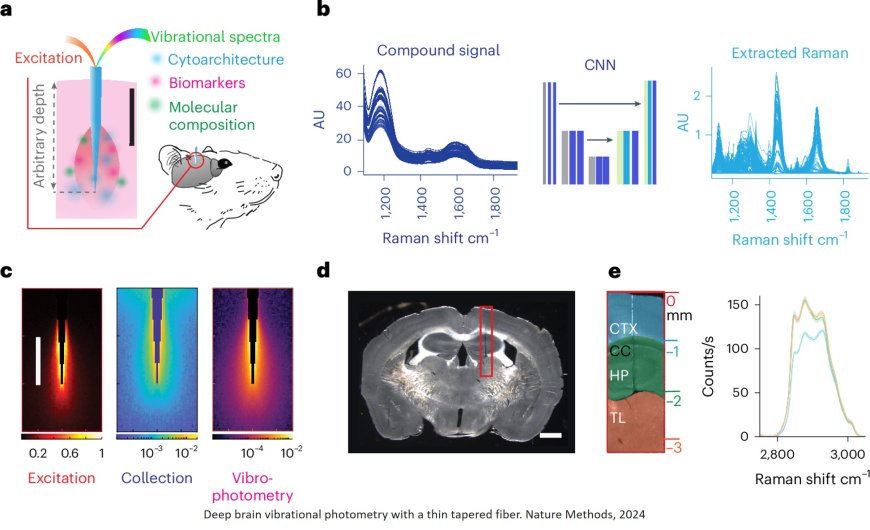
Monitoring the changes caused in the brain at the molecular level by cancer and other neurological pathologies in a non-invasive way is one of the great challenges of biomedical research. A new technique, still in the experimental stage, achieves this by introducing light into the brains of mice using a very thin probe. The innovation, which is published in the journal Nature Methods.
The authors refer to the new technique as a ‘molecular lantern’, as it provides information on the chemical composition of brain tissue in response to light. This allows analysis of molecular changes caused by tumors, whether primary or metastatic, and also by injuries such as head trauma.
The molecular lantern is a probe of less than 1 mm thick, with a tip just a thousandth of a millimetre (a micron), invisible to the naked eye. It can be inserted deep into the brain without causing damage (as an example, a human hair measures between 30 and 50 microns in diameter).
The probe is not yet ready for use in patients; for now it is primarily a ‘promising’ research tool in animal models that allows ‘monitoring molecular alterations caused by traumatic brain injury, as well as detecting diagnostic markers of brain metastasis with high accuracy,’ explain the authors of the paper.
Activating or recording brain function using light is not new. For example, the so-called optogenetic technique make it possible to monitor the activity of individual neurons with light. However, this requires introducing a gene into the neurons that makes them sensitive to light. With the new technology now presented by NanoBright, it is possible to study the brain without altering it beforehand, which represents a paradigm shift in biomedical research.
The new molecular lantern is based on a technique called vibrational spectroscopy, which leverages the Raman effect—a unique property of light. “When light interacts with molecules, it scatters in a way that depends on their composition and chemical structure. This scattering produces a distinct signal, or spectrum, that acts as a molecular fingerprint, providing detailed information about the composition of the illuminated tissue”, explain the author.
“We can see any molecular change produced in the brain by a pathology or injury”
“This technology allows us to study the brain in its natural state; it is not necessary to alter it beforehand. But it also makes it possible to analyse any type of brain structure, not only those that have been genetically marked or altered, as was the case with the technologies used until now. With vibrational spectroscopy we can see any molecular change in the brain when there is a pathology”, explains the author.
Raman spectroscopy is already used in neurosurgery, although in an invasive and less precise way: “There have been studies of its use when operating on brain tumors in patients,’ says the author. In the operating room, once the bulk of the tumor has been surgically removed, it is possible to introduce a Raman spectroscopy probe to assess whether cancer cells remain in the area. That is, it is only used when the brain is already open and the hole is large enough. But these relatively large ‘molecular lanterns’ are incompatible with minimally invasive use in live animal models”.
For the group, one goal now is to find out whether the information provided by the probe allows “differentiating different oncological entities, for example, the types of metastases according to their mutational profiles, by their primary origin or from different types of brain tumours”.
The group has used the technique to investigate the epileptogenic zones surrounding traumatic brain injuries. “We were able to identify different vibrational profiles in the same brain regions susceptible to epileptic seizures, depending on their association with a tumor or trauma. This suggests that the molecular shadows of these areas are affected differently, and can be used to separate different pathological entities by automatic classification algorithms including artificial intelligence”, explains the author.
“The integration of vibrational spectroscopy with other modalities for recording brain activity and advanced computational analysis with artificial intelligence will allow us to identify new high-precision diagnostic markers, which will facilitate the development of advanced neurotechnologies for new biomedical applications”, summarizes the author.
https://www.nature.com/articles/s41592-024-02557-3
https://sciencemission.com/Deep-brain-vibrational-photometry