


Virus-drug structure, interaction in real time
The researchers have uncovered crucial insights into how an emerging class of antiviral drugs works.
The discovery sheds light on an important tool for fighting drug-resistant strains of herpes simplex virus, or HSV, and points to new pathways for treating herpesviruses and other kinds of DNA viruses (those that have DNA as their genetic material and can replicate inside host cells).
The federally funded study is published in Cell.
As an infectious disease doctor, study co-senior author has seen many patients with compromised immune systems develop dangerous drug-resistant HSV infections. Such resistance can evolve when the virus is repeatedly treated with the same type of antiviral.
Members of a new class of antivirals are being tested in U.S. clinical trials, and one has gained approval in Japan, but how these emerging drugs work is mostly unknown. The researchers sought to uncover some of those secrets.
The lab focused on revealing the structural details of how the drugs locked to the viral protein, while another lab worked on capturing real-time details about how the binding process works to block the viral protein.
“A real strength of this study is the combination of high-resolution, atomistic pictures of the viral proteins bound by the inhibitors and real-time imaging of the viral proteins in action,” co-senior author said.
Herpesviruses can cause infections such as chicken pox, shingles, and mononucleosis; have been implicated in cancers, autoimmune diseases, and other diseases; and tend to linger in a latent stage lifelong. Among them, HSV-1 is commonly known for causing cold sores but can also cause serious brain infections in healthy adults and severe disease in immunocompromised people.
The antiviral medicines that are currently FDA-approved for HSV-1 focus on targeting the virus’s DNA polymerase, a protein that makes copies of the viral genome. However, strains of the virus have emerged that are resistant to these drugs.
Alternatives are already on their way, including a class of drugs known as helicase-primase inhibitors, or HPIs. These target the viral helicase-primase, an enzyme that, like the polymerase, is critical for herpesviruses to make copies of themselves.
The viral helicase unwinds the viral genome, motoring along and unzipping the interlaced strands of DNA to convert it to single-stranded DNA. This exposes the information encoded in the genome so the polymerase can reproduce it.
The viral primase, meanwhile, triggers the creation of an RNA molecule that serves as a starting place for the new copy of the genome to attach itself to, like the piece at the bottom of a jacket zipper that allows the slider to engage with the teeth of the zipper.
HPIs interfere with these operations to stop the copying process.
Until now, no research had managed to reveal the structures of HSV enzymes such as the helicase-primase. One reason is that they are very wiggly, constantly moving and changing shape. The existence of a working inhibitor — the HPI drugs — now allows scientists to lock the enzymes into a single, static form that can be imaged. Without it, the authors said, their discoveries wouldn’t have been possible.
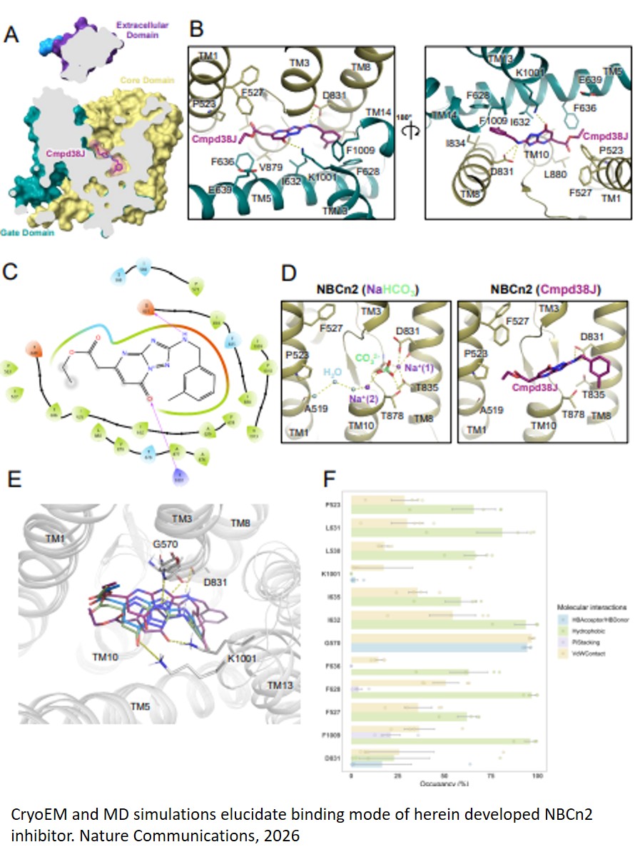
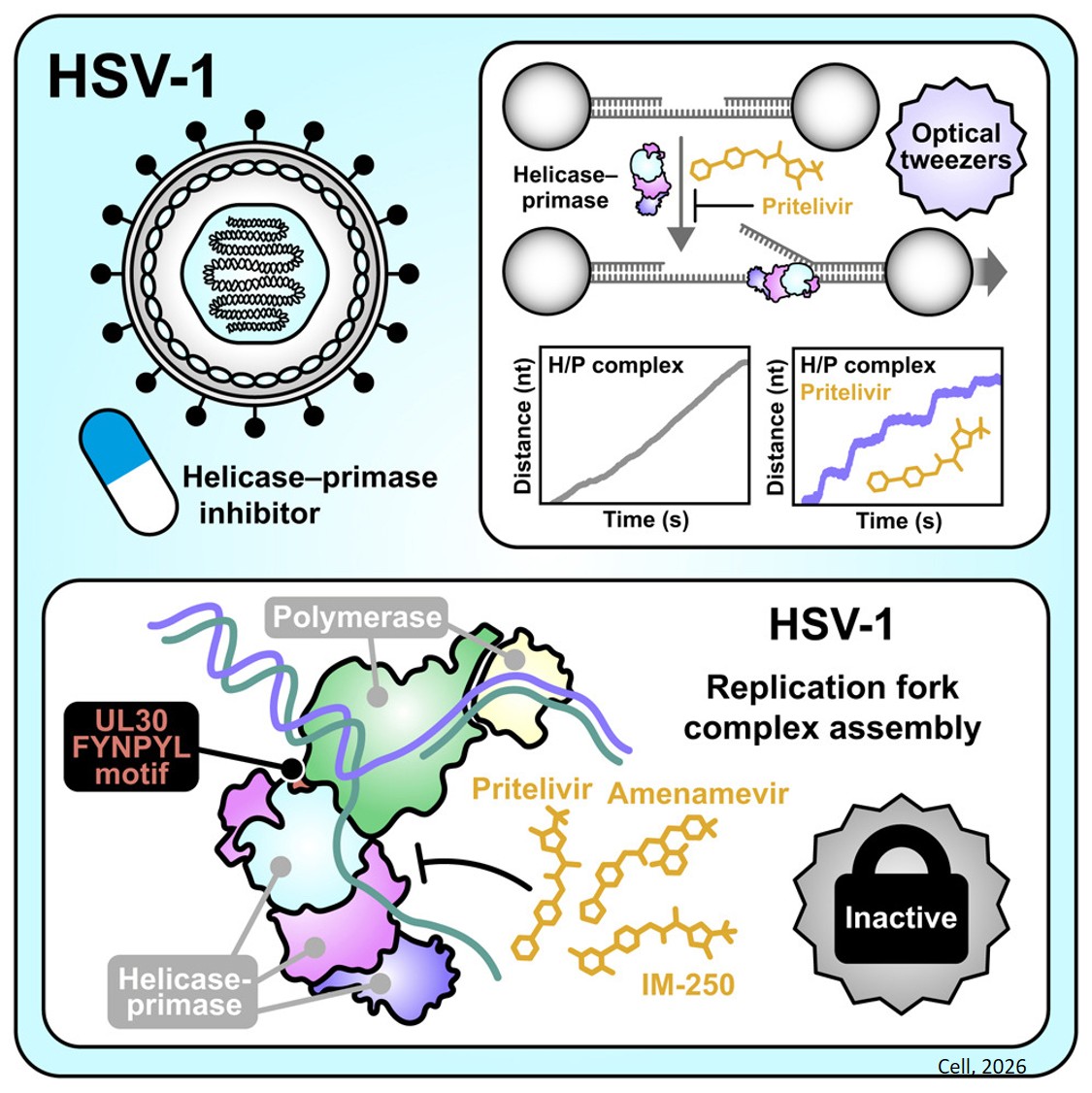
The team used cryogenic electron microscopy (cryo-EM) to visualize at near-atomic resolution the physical structure of the HSV-1 helicase-primase when it is bound by several inhibitors.
The researchers also used cryo-EM to visualize how the viral helicase-primase interacts with the viral polymerase during DNA replication. The structure of this larger complex may help in the identification of new drug target sites by revealing the physical and chemical properties of places where a potential drug could bind and interfere with replication.
A cryo-EM image is a detailed snapshot that shows precisely where and how the drugs interlock with the enzyme and prevent the virus from making new copies of itself. But to understand in a more detailed way how the inhibitor functions, the team wanted to see the molecules in action.
To do this the researchers performed experiments with a tool known as optical tweezers.
Optical tweezers use the momentum of photons from highly focused lasers to manipulate particles, like a science-fiction tractor beam but at a much smaller scale.
The researchers used the tweezers to suspend a piece of viral DNA equipped with the helicase-primase between two beads. They watched the enzyme working, then investigated what happened when they introduced tiny doses of the inhibitor drug. The team was able to observe individual helicase molecules unwinding the double helix of DNA and also could see when the drug stopped the process.
“The latest generation of imaging tools, like the tweezers, have given scientists an unprecedented ability to see how the processes of life work at the level of single molecules,” said the author. “In this case we were able to see, in real time, how the inhibitor gums up the motor of the helicase and causes it to stall.”
These new insights inspire hope that their work can drive further drug development for antivirals and continue to provide deeper knowledge that will someday soon let physicians harness the machinery of life to bring better health to people everywhere, the researchers said.
https://www.cell.com/cell/fulltext/S0092-8674(25)01376-5
https://sciencemission.com/HSV-1-helicase-primase-inhibition