


Characterizing human dermal sleeping pain neurons
Researchers have determined the molecular signature of human sleeping — or silent — nociceptors: sensory neurons that are unresponsive to touch or pressure yet are key culprits in neuropathic pain.
The findings suggest a potential pathway for finding drug targets to relieve chronic pain, said a co-author of the study published in the journal Cell.
“We know from direct human physiological evidence that these cells are important in neuropathic pain,” said the author. “Now we can identify them at the gene-expression level with an astonishing degree of detail. This will allow researchers to start working on targets to manipulate those cells, which could bring about very exciting developments in the future.”
Sleeping nociceptors are a distinct class of sensory neurons that can become spontaneously active to cause persistent pain without an evident stimulus. This makes them essential components of the neuropathic pain suffered by approximately 20% of American adults.
The cell bodies of sleeping nociceptors are located in the dorsal root ganglia, nerve cells clustered near the base of the spine that relay sensory signals from the peripheral nervous system to the central nervous system. The axons and dendrites that protrude from each cell and carry electrical signals are long and connect to the skin. Though the functional properties of these fibers have long been known, their distinctive molecular characteristics remained unclear.
“Active sleeping nociceptors have been found in people with diabetic neuropathy, postherpetic neuralgia and fibromyalgia,” the author said. “There are also scores of neuropathic pain studies that found no cause. If you were going to highlight any type of sensory neuron as the biggest culprit for the spontaneous, shooting pain that neuropathy patients have, it’s these sleeping nociceptors.”
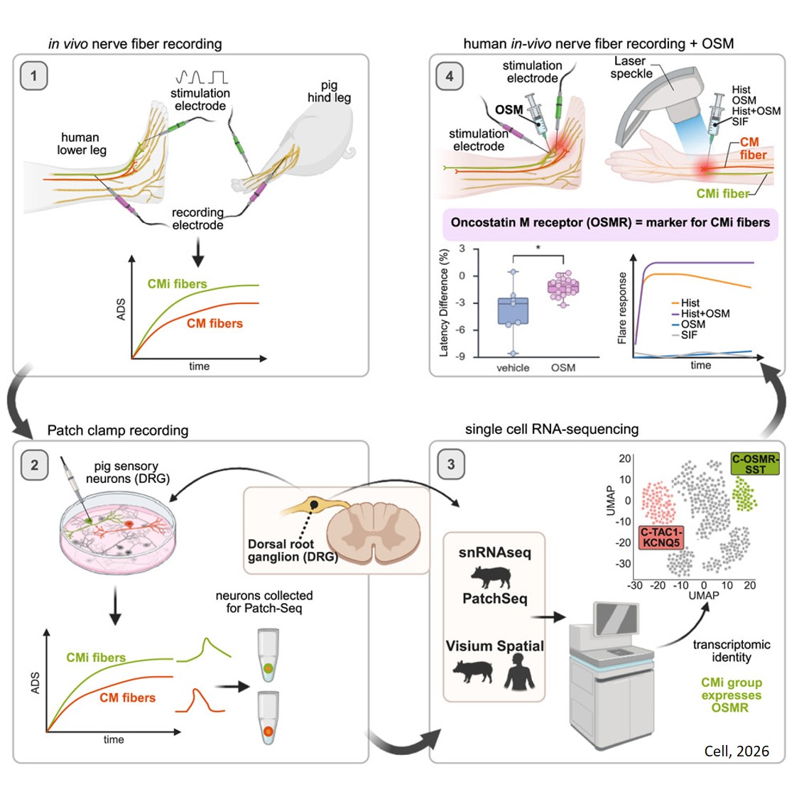
The researchers used high-resolution recordings of electrical activity of individual neurons alongside techniques that read the genetic activity of the neurons to identify sleeping nociceptors among the broader nerve population.
To discover the distinctive molecular profile of the neurons, the researchers first used isolated dorsal root ganglia from pigs because sleeping nociceptors in porcine skin closely resemble those found in humans. Cross-species analyses confirmed that the same molecular markers are present in pig and human sensory neurons. They are both characterized in part by the presence of the oncostatin M receptor on the cell and the neuropeptide somatostatin, which suppresses the release of various hormones.
“Our work establishes a new conceptual framework for understanding the emergence of neuropathic pain at the molecular level, opening concrete perspectives for the development of targeted therapies,” the author said.