


Childhood brain diseases from mutations in tubulin co-factors
Thousands of times per year, a family’s moment of joy turns to unexpected grief. A seemingly healthy infant stops smiling or making eye contact. Their limbs grow weak. The tiny child suffers seizures and breathing problems. Their child has a rare genetic disorder called a chaperone tubulinopathy.
“The parents are asking if there’s a way to do gene therapy,” the senior author said. These life-shortening diseases, with names like infantile encephalopathy, corpus callosum hypoplasia and Kenny-Caffey syndrome, currently have no treatments. But the research team have made a major discovery that could change that.
In two scientific papers, they have mapped the structure and mechanics of a critical cellular machine that malfunctions in people with these diseases.
In addition to enabling new treatments, this discovery could help scientists identify dozens of other genetic diseases in which children experience various neurological problems with no clear explanation.
The researchers study structures called microtubules, which form protein skeletons inside cells. As a cell grows and changes shape, its cytoskeleton drives the process – lengthening its scaffold of microtubules.
“They are the cell’s force generators,” the author said. These telescoping structures are crucial in the developing nervous system.
They help nerve cells grow the long tendrils, called axons, that connect with other nerve cells. These axons allow neurons to communicate across long distances — especially in the optic nerves that connect the eyes to the brain, in the corpus callosum that connects the left and right hemispheres of the brain, and in the long nerves that reach down to the arms, legs, lungs and other organs.
For a baby to develop normal vision, cognition, coordination and breathing, the neurons have to connect properly. The microtubules must form perfectly inside the growing neurons.
The cell builds microtubules from two proteins, called α-tubulin and β-tubulin. Before they can be used, they have to be snapped together into thousands of αβ-tubulin “dimers” – forming the building blocks that can then assemble into microtubules.
Cells control the formation of microtubules, in part, by controlling the supply of the αβ-tubulin dimers. Special “chaperone” proteins – called “tubulin cofactors” – perform this delicate process. As soon as a cell produces a β-tubulin protein, these cofactors assemble into a cage that holds onto the β-tubulin until it can find an α-tubulin and snap them together into an αβ heterodimer, which it then releases.
But this process can easily go awry, with terrible consequences.
If the tubulin cofactors malfunction, it reduces the cell’s supply of αβ-tubulin, disrupting the microtubules that guide neuronal growth. “Even a small percent decrease in αβ-tubulin supply is toxic to the cell,” the author said.
Scientists have discovered that some children with severe, unexplained neurologic disorders actually have mutations in their tubulin cofactor genes. This may reduce the supply of αβ-tubulin – leading to underdeveloped corpus callosum, optic nerves and other brain structures.
“Some of these mutants were identified almost 35 years ago in yeast,” the author said. They were discovered 15 years later in humans. But the delicate proteins were difficult to study, the author said, “so this whole field of research was essentially shelved for a long time.”
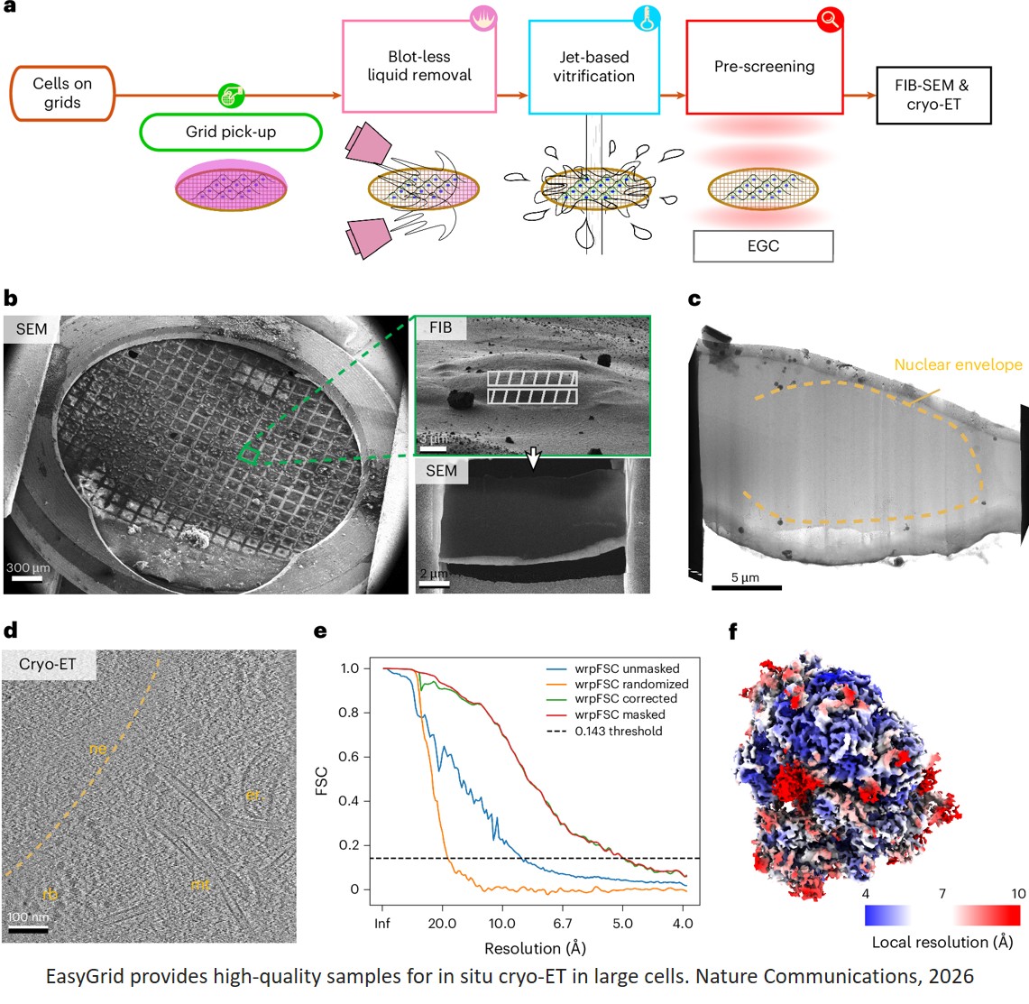
Using cryo-electron microscopy (Cryo-EM), they found how these proteins assemble into a complex machine. Their initial results, published in Nature Communications, show the elegant spring-and-latch mechanism that it uses to capture β-tubulin, snap it onto α-tubulin, and release the αβ dimer. “This was a surprise,” the author said. “It was really beautiful.”
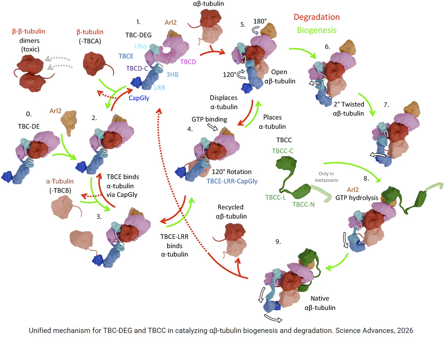
In the second paper, published in Science Advances, the authors unveil additional cryo-EM structures, showing the machine frozen in at least nine different configurations. These snapshots reveal how it functions in a complex cycle, snapping together αβ-dimers when they are needed, and pulling them apart when they aren’t.
The tubulin cofactors, TBCC, TBCD, and TBCE and the Arl2 GTPase, form TBC-DEG assemblies that regulate the assembly of α- and β-tubulin into heterodimers and their disassembly.
The structures reveal that TBC-DEG disassembles αβ-tubulin by releasing α-tubulin through a lever arm–like rotation in TBCE coupled to major conformational change in Arl2 upon its nucleotide release, while TBCD tightly holds β-tubulin. TBCD dissociates α-tubulin by refolding the β-tubulin H10-S8 loop at its intradimer interface.
The TBC-DEG–β-tubulin or TBC-DE–β-tubulin assemblies undergo extensive back-to-back dimerization mediated by β-β-tubulin homodimers, formed through their dissociated H8 helices at unoccupied intradimer interfaces.
Structural comparisons demonstrate that TBCE’s mechanical rotation, driven by the Arl2 GTPase cycle, either delivers α-tubulin or removes it from beneath the TBCD-bound β-tubulin and is directionally regulated by TBCC stabilizing αβ-tubulin interfaces.
These discoveries won’t immediately lead to treatments, but they could offer hope to affected families, the author said: “For the first time, we have a precise picture of exactly what’s going wrong, and what a future therapy would need to fix.”
They could also allow disorders to be diagnosed more quickly. Today, families often endure a diagnostic odyssey in which the genomes of parent and child are sequenced in search of a mutation that might explain the problem – often yielding inconclusive results. A clearer understanding of which mutations disrupt the function of tubulin cofactors could lead to quicker diagnosis.
This new knowledge might even spur the discovery of other genetic disorders still flying under the radar.
“Many children are born with minor, unexplained neurologic disorders,” the author said. “Some of them may turn out to have small changes in these genes. Finding that out would be a huge step forward.”
https://www.nature.com/articles/s41467-025-68142-0
https://www.science.org/doi/10.1126/sciadv.aee2303
https://sciencemission.com/Cryo-EM-structures-of-the-tubulin-cofactors