


Brain probe powerfully records neural circuits during behavior
Trying to document how single brain cells participate in networks that govern behavior is a daunting task. Brain probes called Neuropixels, which feature high-density silicon arrays, have enabled scientists to collect electrophysiological data of this nature from a variety of animals. These include fish, reptiles, rodents and primates, as well as humans.
Neuropixels, which come in several versions, record electrical activity from hundreds to thousands of neurons simultaneously. Neurons are nerve cells that receive, process and transmit information.
While the data collected has led to insights on the neural basis of perception and decision-making, those probes cannot sample fine-scale brain structures. They also are limited in resolving (separately distinguishing) the electrical fields around individual brain cells.
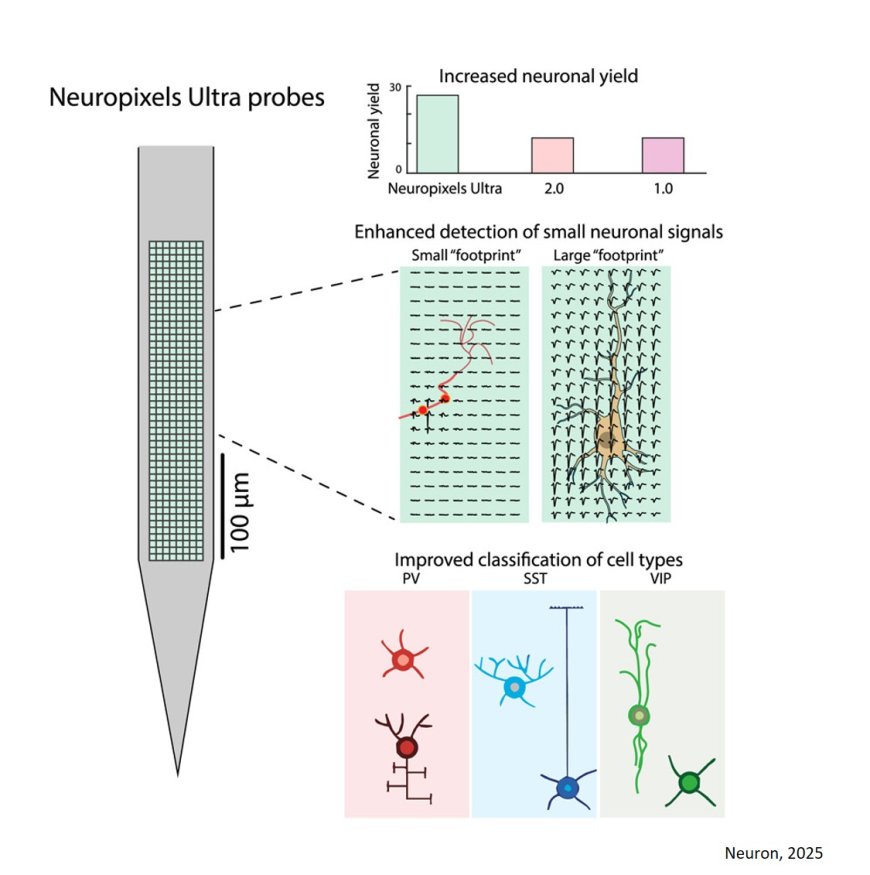
A newly developed probe, called Neuropixels Ultra, overcomes some key technical challenges in recording the cell type and activity of thousands of individual cells across many brain regions during a single experiment.
The probe is essentially an implantable, voltage-sensing camera that can capture flat images of a brain cell’s electrical field.
“We developed a silicon probe with much smaller and denser recording sites than previous designs,” said the corresponding author of a report on tests of the probe’s function.
Their paper appeared in the journal, Neuron.
By obtaining more detailed information about the electrical field surrounding a brain cell than was previously possible, the new probe improved scientists’ ability to detect and classify individual cells.
For example, the researchers conducted recordings of the mouse visual cortex — an area near the back of the brain that processes and interprets information coming from the eyes. The scientists were able to detect twice as many brain cells, compared with recordings with other Neuropixels versions. They also could distinguish three cortical cell subtypes from each other and from other neurons. Being able to detect specific cell subtypes is important in studying brain circuits.
Because the probe gathered data from smaller sites, it predictably had higher noise levels per channel than do earlier probes. But because it sampled 10 times more recording sites, it still offered better data quality.
Neuropixels Ultra was more precise in estimating the spatial position of spikes, the rapid rise and sudden fall of an electrical impulse from a brain cell. It was better at separating spikes from one neuron and not attributing them to nearby neurons. This spatial precision helped increase the yield of sortable, visually responsive neurons. Such measurements could help scientists more accurately decode and track brain cell performance related to visual stimuli.
The researchers also used Neuropixels Ultra to determine how prevalent small-footprint extracellular spikes were across different brain regions and in species other than mice. Thes animals included electric fish, bearded dragon lizards and pigtailed macaques.
“These small spatial footprints, which are difficult to detect with lower density probes, are consistently detected with Neuropixels Ultra,” the scientists observed. They also found that the spatial footprints of cell types that were genetically identified were different for each cell type. This information could help, for example, in determining which cells are present and their distinctive electrical activity.
The researchers concluded that there were tradeoffs between the newer and older probes, depending on the type of experiments a lab conducts. Although the newer probe has a smaller site size and spacing, it also has what its developers describe as “unprecedented” resolution. By contrast, the older probes have a larger vertical recording span. Nonetheless, the recent findings “highlight the advantages of electrophysiological probes with increased site density for a wide range of neurosciences applications,” the researchers noted.