How calcium signals help cells bury their dead neighbors
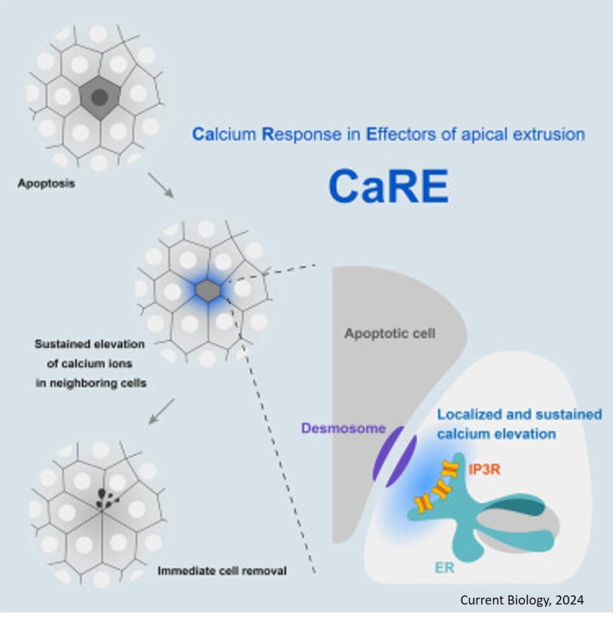
A research team has recently discovered a calcium-based mechanism that plays a key role in the disposal of dead cells, shedding light on how our bodies protect themselves from injury and disease. In their study, published in Current Biology, the team unveiled how calcium ion levels are essential for the efficient removal of dying or apoptotic cells from epithelial tissues (cells lining the body surface), using genetically engineered epithelial tissue cultures, molecular markers, and advanced imaging techniques.
The surfaces of our bodies, including the skin and internal organs, are covered by sheets of epithelial cells that act as vital barriers. When these cells become damaged and die (apoptosis), neighboring cells quickly band together to push them out and seal any gaps through which foreign substances could enter and potentially lead to infections or inflammation. Although this complex process is essential for maintaining a healthy epithelial barrier, the exact mechanism underlying it has not been entirely clear—until now.
To begin with, the team induced apoptosis in individual epithelial cells using a focused laser and observed the response in the surrounding cells. They then observed how nearby cells reacted by modifying them to express special calcium ion probes called GCaMP6, which allowed them to visualize real-time calcium changes. Interestingly, they found that the neighbors of the apoptotic cell showed a significant spike in calcium levels, particularly near the membrane regions interfacing with the dying cell. The researchers named this intriguing phenomenon the “calcium response in effectors of apical extrusion (CaRE).”
Delving deeper into this newly discovered mechanism, the team next examined the role of IP3 receptors, proteins present inside cells that help regulate calcium ion levels. They found that inhibiting the activity of IP3 receptors or removing their associated genes completely prevented the expulsion of apoptotic cells. Further analysis using advanced electron microscopy revealed that a specific subset of IP3 receptors, particularly those located near desmosomes, plays a key role in CaRE.
Desmosomes are cell adhesion structures that form strong connections between cells, acting like buttons that hold them together. They are especially important in tissues like skin and organ linings, helping to keep everything intact and functioning properly. By ensuring neighboring cells adhere tightly, desmosomes play a key role in maintaining the structure and stability of our body’s tissues. The team found that the activation of IP3 receptors near desmosomes is necessary for triggering the contraction of a group of proteins known as actomyosin complex, which helps cells change shape and move, facilitating the removal of apoptotic cells.
“Our study sheds light on a newfound role of IP3 receptors in desmosomes, the latter of which were previously thought to be involved only in mechanical connections between epithelial cells,” highlights the author.
As this study was conducted on cultured cells, the team notes that further analysis of the CaRE mechanism are needed to determine whether the mechanism also function in living organisms, if it varies between different organ tissues, and whether other factors also play a role.
Overall, this study advances our understanding of how our bodies maintain a healthy epithelium—something many of us take for granted. “Our findings provide valuable insights into understanding diseases caused by epithelial barrier disruption, such as atopic dermatitis and inflammatory bowel disease, and may contribute to the development of new preventive measures and treatments for chronic inflammation,” concludes the author.
https://www.cell.com/current-biology/fulltext/S0960-9822(24)01173-4
