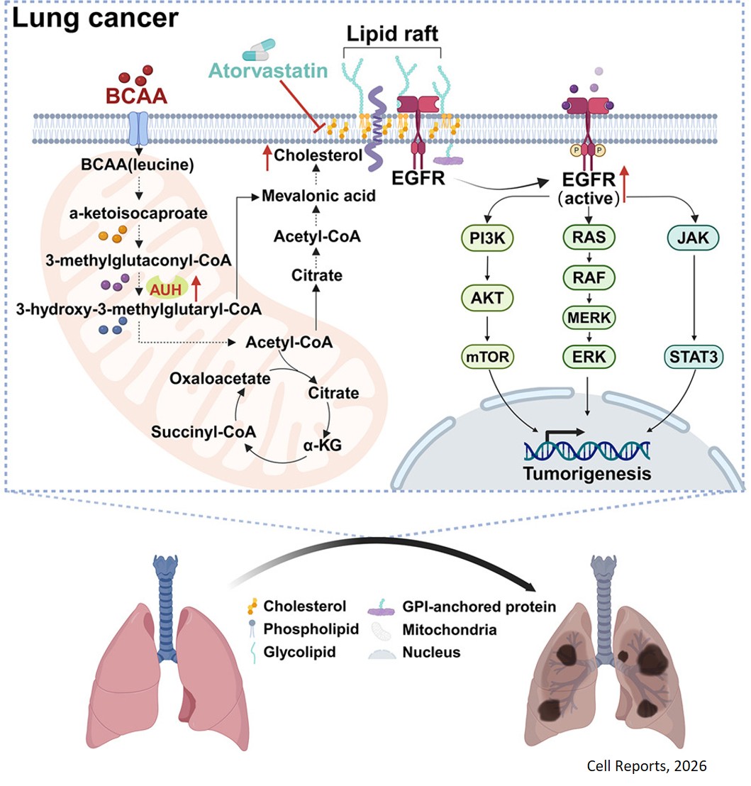


New method reveals structures of RNA-protein complexes in living cells

A new method developed allows researchers to better understand how RNA works. The method, published in Molecular Cell, is a powerful strategy for identifying intricate RNA structures that are likely to have significant biological functions inside cells.
“RNA molecules perform many functions inside cells. While RNA is mostly known for its role as a messenger that carries genetic instructions from DNA to make proteins, RNAs also coordinate and regulate many other cellular processes, and they accomplish this by binding proteins and other molecules,” said the corresponding author.
To carry out its functions, RNA naturally folds into complex, highly dynamic 3D shapes, forming pockets, bumps and knots that interact with specific molecules in the cell. In many cases, RNA structures cycle between different conformations with molecular interactions that support distinct functional outcomes.
“An important step to better understand cellular processes involving RNA is to be able to define RNA 3D structures that are key to those processes, as well as their dynamics,” the author said. “Current biochemical approaches for identifying RNA structures that bind to specific proteins or drugs are complex and laborious and sometimes do not provide answers. We have developed an innovative, one step biochemical approach called multi-site DMS-MaP (msDMS-MaP) that allows us to identify RNA 3D structures and protein binding sites that are crucial to specific cell functions.”
For instance, the researchers applied msDMS-MaP to answer a 50-year-old question in the field of biochemistry – how does RNA folding influence the assembly of ribosomes, the cellular machinery that synthesizes proteins? Using bacterial ribosomes as a model, they discovered that ribosomal RNAs encode numerous independently folding 3D structures that coincide with binding sites for proteins that drive the assembly of ribosomes.
The new method offers several advantages when compared to alternative methods for probing RNA 3D structure: experiments are easy to perform, can directly measure RNA folding in cells, require only inexpensive and widely available reagents and are compatible with most high-throughput protocols, making it possible to scale the method to study thousands of RNAs at once.
“We found that msDMS-MaP is a powerful strategy for resolving RNA 3D structures and protein binding sites that are normally hidden to existing methods. Having this information helps us better understand how RNAs work in normal cellular processes and how things go wrong in disease. In the future, the method also could be applied to develop and optimize RNA-targeting drugs and RNA vaccines,” the author said.
https://www.cell.com/molecular-cell/fulltext/S1097-2765(26)00205-4