


Telomeres and centromeres interact in some aggressive cancers
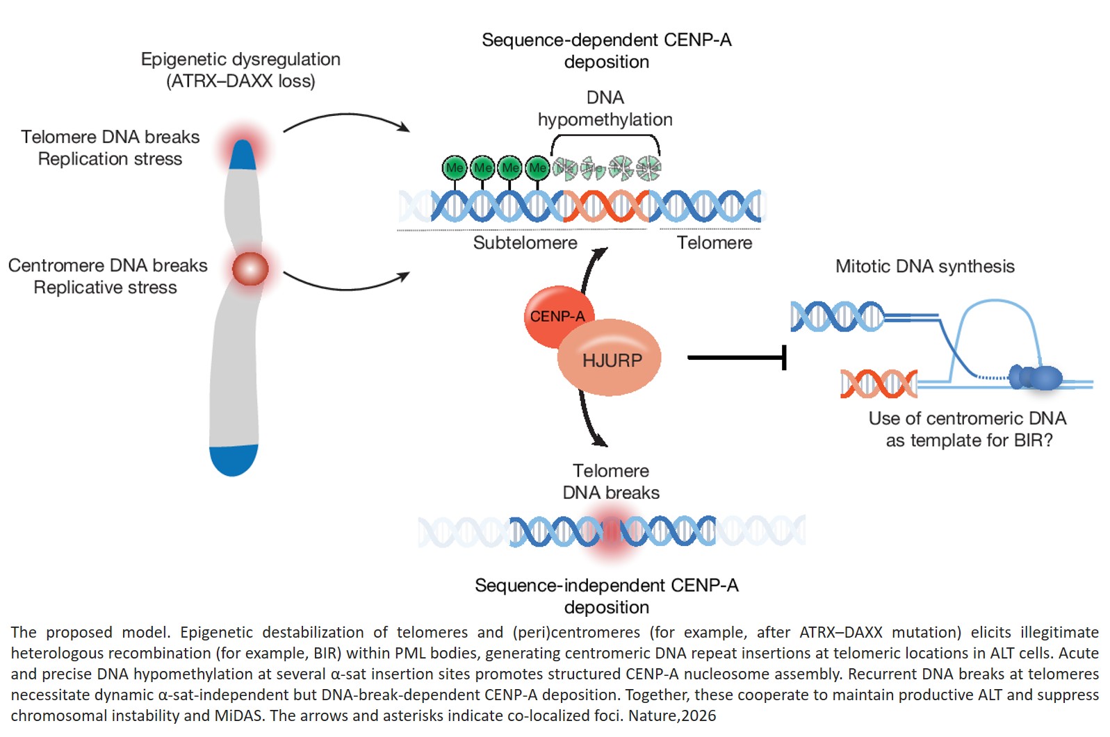
Published in Nature, researchers report a previously unrecognized change in how the cell’s genetic material is packaged into structures called chromosomes, that helps explain how some aggressive cancers sustain unlimited growth.
In a subset of tumors known as alternative lengthening of telomeres (ALT)-positive (ALT+) cancers, the team found that DNA normally associated with centromeres—the central regions of chromosomes—can be inserted near telomeres, the chromosome ends. Because this pattern appears in patient tumors, including pediatric brain cancers, it could serve as a biomarker to identify tumors driven by these unusual genetic rearrangements and track their evolution over time.
“This is something that nobody expected. These are two parts of the chromosome that are never supposed to interact,” said a senior author. “It is not just interesting biology, but it tells us something fundamental about ALT tumors,” said the author.
For decades, scientists have understood that DNA is packaged into chromosomes organized into distinct functional regions. At chromosome ends, telomeres protect genetic material, while centromeres act as anchors that ensure chromosomes are properly separated during cell division. Because of their distinct roles, they have been thought to remain separate, with their strict separation considered essential for maintaining genome stability. The new findings show that this organization can break down in cancer cells, allowing these regions to interact in ways previously thought impossible.
The work focused on a subset of cancers known as ALT+ tumors, which use a mechanism called alternative lengthening of telomeres (ALT) to maintain chromosome ends. This process allows tumor cells to maintain telomere integrity and continue dividing without relying on telomerase, the enzyme most cells use for this function. Found in approximately 5–10% of cancers overall, ALT occurs in a subset of tumors within several cancer types, including pediatric neuroblastoma, and is often associated with genomic instability, which makes these cancers particularly challenging to treat. Despite decades of research, however, the specific structural changes in the genome that enable ALT to function have remained unclear—until now.
In both laboratory models and real tumors, ALT-positive cancers showed higher levels of mixed, or chimeric, centromere and telomere DNA than ALT-negative tumors, supporting the idea that this is a defining feature of these cancers rather than an arbitrary defect. The study also found that, for ALT+ cells to acquire centromere-like features at their telomeres, they depend on underlying epigenetic changes—alterations in how DNA is packaged and regulated— including the loss of a chromatin regulator called ATRX, which normally helps keep these regions separate. When the researchers disrupted this process, telomeres became unstable and reduced ALT activity.
“It is remarkable that the illegitimate recombination between centromere and telomere sequences, which may begin as a mistake inside the cell, is actually being used by cancer cells to adapt and survive,” explains a co-corresponding author of the study.
The findings also point to clinical relevance, particularly for cancers in which ALT is prevalent. The centromere–telomere signature identified in this study offers a potential molecular marker for distinguishing ALT-driven tumors and for better understanding how they progress. By revealing a structural feature that appears unique to these cancers, the work provides a foundation for identifying patients, monitoring disease evolution and exploring new therapeutic strategies.