Human assembloid model of the ascending neural sensory pathway

Investigators have replicated, in a lab dish, one of humans’ most prominent nervous pathways for sensing pain. This nerve circuit transmits sensations from the body’s skin to the brain. Once further processed in the brain, these signals will translate into our subjective experience, including the uncomfortable feeling of pain.
The advance promises to accelerate what has been slow progress in understanding how pain signals are processed in humans and how best to alleviate pain.
In a study published in Nature, scientists describe their successful assembly of four miniaturized parts of the human nervous system to reconstitute what’s known as the ascending sensory pathway.
The peripheral sensation of pain travels to the brain in a relay involving nerve cells, or neurons, centered in four different regions of the ascending sensory pathway: the dorsal root ganglion, dorsal spinal cord, thalamus and somatosensory cortex.
“We can now model this pathway non-invasively,” said the study’s senior author. “That will, we hope, help us learn how to better treat pain disorders.”
Until now nobody has been able to watch information being transmitted through this entire pathway. But the authors witnessed never-before-seen waves of electrical activity travel from the first component of their construct all the way to the last, and they were able to enhance or disrupt these wavelike patterns by gene alterations or chemical stimulation of elements of the circuit.
The regions that compose the ascending sensory pathway are linked by three sets of neuronal connections: The first set relays sensory information from the skin through the dorsal root ganglion to the spinal cord; a second set of neurons passes the signals from the spinal cord to a brain structure called the thalamus; and the third relays this information from the thalamus to the somatosensory cortex for further processing of the signal originating from the periphery.
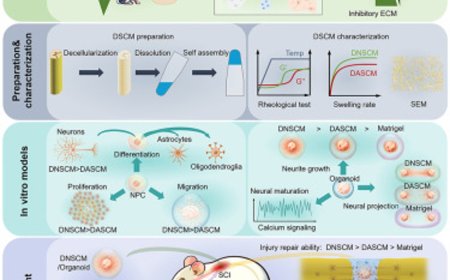
In the new study, the authors developed human organoids recapitulating the ascending sensory pathway’s four key regions, then fused them together in series to form an assembloid mimicking the pathway. Starting with cells from skin samples from volunteers, the team first transformed them into induced pluripotent stem cells, which are essentially de-differentiated cells that can be guided to become virtually any cell type in the human body. The researchers used chemical signals to coax these cells into aggregating into tiny balls, called neural organoids, representing each of the four regions of the pathway.
Each organoid was a bit less than 1/10 inch in diameter and contained close to a million cells.
The authors lined up the organoids of those four different types side by side and waited. About 100 days later, they had fused into an assembloid almost 2/5 of an inch long — “They look like tiny sausage links,” the author said — and consisting of nearly 4 million cells.
That’s less than 1/42,000th of the number in an adult human brain, which contains about 170 billion cells, Pasca noted. But the construct recapitulated the circuitry involved in the pathway.
The researchers showed that the assembloids’ constituent organoids were anatomically connected: Neurons from the first had formed working connections with neurons from the second, the second with the third and so on.
Moreover, the entire circuit, from the sensory organoid to the cortical organoid, worked as a unit. Once all four organoids had sat beside one another in the flask for about 100 days, patterns of spontaneous, synchronized, directional signaling within the assembloid began to emerge: Neuronal activity in the sensory organoid tripped off similar action in the spinal organoid, then in the thalamic organoid and finally in the cortical organoid.
“You’d never have been able to see this wavelike synchrony if you couldn’t watch all four organoids, connected, simultaneously,” the author said. “The brain is more than the sum of its parts.”
Chemicals known to induce pain increased the wavelike activity in the assembloids. Stimulating the sensory organoid with capsaicin — the ingredient in chili peppers that produces a burning sensation in our mouths — triggered immediate waves of neuronal activity.
Rare genetic mutations in an ion-exchange protein found on the surfaces of peripheral sensory neurons can lead to debilitating hypersensitivity to pain or, conversely, a life-threatening inability to experience pain — radically increasing the physical dangers, routine or otherwise, that life serves up.
The protein in question, Nav1.7, is a particular type of sodium channel that, the researchers observed, abounds on peripheral sensory neurons but is scarce elsewhere. The scientists made an assembloid with its initial, sensory component’s normal version of Nav1.7 replaced by the mutant pain-hypersensitivity version. The resulting mutant sensory assembloids displayed more-frequent waves of spontaneous nervous transmission from the sensory through the spinal and thalamic organoids to the cerebral-cortex organoid.
When the team instead rendered the same sodium channel in the sensory organoid non-functional, they got a surprise: Firing from that organoid in response to a pain-inducing chemical continued — but the synchronized wavelike transmission of pain information through the circuit mysteriously vanished. The firing was now out of sync.
“The sensory neurons still fired,” the author said. “But they failed to engage the rest of the network in a coordinated manner.”
Missing in these assembloids, importantly, were organoids representing other brain regions that are critical to the discomfort experienced by people who are in pain.
“The assembloids themselves don’t ‘feel’ any pain,” the author said. “They transmit nervous signals that need to be further processed by other centers in our brains for us to experience the unpleasant, aversive feeling of pain.”
The assembloids, after only a few months of their assembly, represent an early phase of fetal development, the author said. Their immediate use would be in studying neurodevelopmental disorders such as autism. People with autism are often hypersensitive to pain and to sensory stimulation in general, and some autism-associated genes are active in the sensory neurons of the ascending sensory pathway.
“Nav1.7 appears to exist mostly on the surfaces of peripheral pain-sensing neurons,” the author said. “We think screening for drugs that tame sensory organoids’ ability to trigger excessive or inappropriate waves of neuronal transmission through our assembloid, without affecting the brain’s reward circuitry as opioid drugs do — which is why they’re addictive — could lead to better-targeted therapies for pain.”
