


How the body senses cold
When you reach into a bucket of ice, open your front door on a snowy day, or feel the tingle of menthol toothpaste, a protein in your nerve cells called TRPM8 springs into action, opening like a tiny gate to send a “cold” signal to your brain.
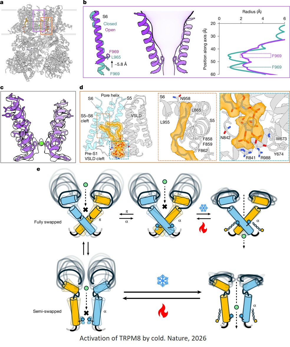
Now, researchers have discovered how TRPM8 changes its shape when exposed to cool temperatures. The work, published in Nature could one day be used to help treat pain that is triggered by cold. It also answers a long-standing question about why birds — which also have TRPM8 in their nerve cells — are far less cold sensitive than mammals.
“Everyone always wants to know how temperature sensing works, but it turns out to be a very technically challenging question to answer,” said co-senior author. “So, to finally have insight into this is really very exciting.”
“For decades, structural biology has focused on capturing proteins in stable, frozen states. This work shows that to truly understand how a protein functions, you also have to understand how it moves,” added co-lead of the work.
Scientists knew that TRPM8 only begins to activate when temperatures dip below about 79 degrees Fahrenheit — and that it was responsible for both cold sensation and the cool feeling of menthol. Yet despite years of effort, researchers had been unable to capture its exact molecular structure while responding to cold.
TRPM8 is normally found embedded in the outer membrane of nerve cells and tended to fall apart when researchers isolated it. Most imaging methods also rely on proteins being locked in a single, stable structure to visualize them — limiting scientists’ ability to see fluid, intermediate structures as a protein changes shape.
The researchers solved this by imaging TRPM8 while it was still embedded in membranes that were taken directly from cells.
“We realized that the protein is particularly sensitive to how you handle it. Keeping it in the native membrane was what finally let us see what was actually happening,” said a co-first author of the study.
To capture what was happening as TRPM8 opened, the team used two complementary techniques: cryo-electron microscopy (cryo-EM), which takes static pictures, and hydrogen-deuterium exchange mass spectrometry (HDX-MS), which is more dynamic.
For cryo-EM, they prepared samples of the protein in cold, with menthol, or at room temperature. Then, they flash froze the samples. This locked the channel into its configuration at that moment. Cryo-EM then generated three-dimensional snapshots of the protein's atomic arrangement.
They used HDX-MS to track the protein in real time as the surrounding temperature changed. The method highlighted which regions of the molecule flex and move as the temperature changed. Together, the methods let the researchers model exactly how TRPM8 opened below 79 degrees.
“Just as looking at a photo of a horse can’t tell you how fast it runs, the electron microscopy alone can’t tell us how the molecule moves and what drives those movements,” said another co-first author. “But combining these two techniques gave us a window into what was happening.”
The analysis revealed that cold stabilizes a specific region of the TRPM8 channel, which then triggers a key helix to move. This enables a separate lipid molecule to slide into that spot, locking the channel open and sustaining the cold signal. When the researchers compared human TRPM8 with the bird version of the protein, which responds to menthol but is far less cold-sensitive, they were able to detect which features are specifically responsible for detecting cold.
The new work paves the way for determining the structure of other dynamic proteins that have typically been hard to image.
“The lessons we learned in studying this channel are actually very broadly useful,” the author said. “Dynamic behavior is critical for the function of many proteins, and you can’t understand dynamic behavior from one snapshot of a protein’s structure.”
The researchers also plan to examine how compounds that block TRPM8 — several of which are in clinical trials for pain — affect the structure of the protein. That could ultimately contribute to more targeted treatments for conditions like cold allodynia, in which even mild cold triggers severe pain.