




How critical brain cells misfire in Alzheimer's
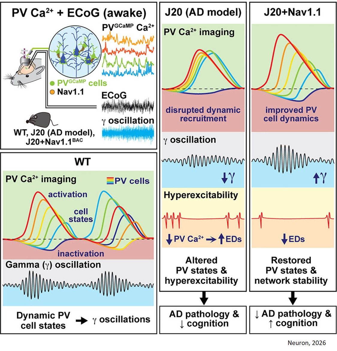
Whether focusing on a task, learning a new skill, or retrieving a cherished memory, the human brain relies on rapid, rhythmic electrical pulses known as gamma oscillations. For decades, scientists knew these rhythms were weakened in Alzheimer’s disease. But how individual brain cells coordinate their activities to generate them—and why that process fails—remained a mystery.
Now, researchers have unlocked a key piece of the puzzle.
In a study published in Neuron, scientists discovered that specialized rhythm-keeping cells called parvalbumin (PV) interneurons generate these brain rhythms by taking precise turns firing. But in mouse models of Alzheimer's disease, this delicate coordination collapses, causing memory waves to falter.
Critically, by boosting levels of a specific protein that helps these PV cells generate electrical pulses, the researchers were able to restore the normal rhythm and brain activity, offering a promising therapeutic target for restoring cognitive function.
“Gamma oscillations are essential for clear thinking and memory, but we've lacked a complete picture of how the brain creates them,” says a senior author of the study. “What makes this discovery so exciting is that we didn't just figure out how healthy cells pass the baton; we discovered a way to reset their timing when Alzheimer's related processes throw them off beat.”
Gamma oscillations are fast, repetitive brain rhythms—cycling 30 to 90 times a second—that happen in the brain’s background during tasks requiring attention and memory. Previously, scientists relied on methods like optogenetics to activate all PV interneurons simultaneously to demonstrate that PV cells generate gamma rhythms. However, broad stimulation couldn't reveal how individual cells behave naturally in real time.
“Turning on all the cells at once shows you what they can do, but not how they naturally work together or how they’re recruited to generate spontaneous gamma oscillations,” says the first author. “Until now, no one has actually been able to image these cells and look at gamma oscillations at the same time in an awake animal during natural behavior.”
To overcome this obstacle, scientists developed an innovative technique combining real-time electrical recording with imaging of live cells. By monitoring mice as they ran on a treadmill—a state that naturally prompts attention-related brain rhythms—the researchers engineered PV cells to glow fluorescently every time they fired an electrical pulse.
The images revealed an intricate relay race: every time a burst of gamma oscillations began, PV cells became active not all at once, but in successive waves. The first cells to respond showed high activity, followed by subsequent groups with progressively weaker firing, while roughly a third of the cells remained quiet—demonstrating a highly organized pattern of turn-taking essential for healthy brain function.
“The relationship between the PV cells and the gamma oscillations was much more dynamic and complex than we expected going into this,” the author says. “Rather than all the cells contributing to the rhythm at once, each cell contributes for just a brief moment, and those small contributions add up to create the larger brain rhythm. It’s less like a military lockstep march and more like a flock of birds in the sky—each responding to the others, shifting turns and trajectories, yet together creating a remarkably coordinated pattern.”
The team then studied mice engineered to develop Alzheimer’s-like disease. In these mice, the rhythm’s timing broke down; PV cells that fired first were sluggish, those that joined late were overactive, and fewer cells stayed quiet. This resulted in too much synchrony—more cells fired at the same time and level, and gamma rhythms became weaker.
“The important thing here is that more activity is not always going to produce better oscillations or better cognition,” the author says. “The dynamics of the cells are most important.”
The findings could help explain why more than a quarter of people with Alzheimer’s also show epilepsy-like activity in the brain. In the mice involved in the study, epileptic spikes appeared after sudden drops in PV interneuron activity.
The lab previously found that levels of Nav1.1—a sodium channel found on the surface of PV cells that helps them fire—are reduced in both Alzheimer’s mouse models and people with the disease. In the new study, the team engineered the Alzheimer’s mice to produce more Nav1.1 and then watched their PV cells and brain activity.
The extra channels restored normal waves of PV interneuron activity and strengthened gamma oscillations. They also reduced the epileptic activity, reduced amyloid plaques (a pathological hallmark of Alzheimer’s), and improved the memory, cognition, and lifespans of the mice. More work is needed to understand the link between PV cell failure and Alzheimer’s in humans, and how Nav1.1 levels or activities could be boosted.
“We still don’t know why interneuron dysfunction happens early in Alzheimer’s disease," the author says. “But this is another study underscoring that a lot of the pathology of the disease could arise downstream of disrupted interneurons and the abnormal brain rhythms this disruption produces.”
“One of the most exciting aspects of this work is our ability to target a single cell type out of the hundreds in the brain,” another author says. “Because these tools work in both mice and humans, they help us bridge the gap between understanding how individual cells fail in disease and developing targeted treatments to fix them.”