


The relationship between brain cellular senescence and brain structure
Researchers have characterized how cellular senescence—a biological process in which aging cells change how they function—is associated with human brain structure in both development and late life. The study, published in Cell, provides new insight into how molecular signatures of cellular senescence that are present during development and aging mirror those associated with brain volume and cortical organization.
Understanding brain structure is a central challenge in neuroscience. Although brain structure changes throughout life and is linked to both aging and neurodegenerative conditions such as Parkinson’s and Alzheimer’s diseases, the underlying molecular processes involved—including cellular senescence—are not defined. Cellular senescence is commonly defined as a state characterized by permanent cell cycle arrest in the absence of cell death, in which cells have altered function. While cellular senescence has been implicated in aging and disease, its role in shaping human brain structure—both during development and aging—has remained unclear.
“This is the first study to directly link senescence-related molecular networks in living human brain tissue to measurable differences in brain structure within the same individuals,” said a co-senior author of the paper. “By identifying molecular pathways that are engaged in both brain structure development and aging, our work highlights senescence as a fundamental biological feature of brain aging and neurodegenerative disease and helps prioritize targets for future experimental research aimed at protecting brain health.”
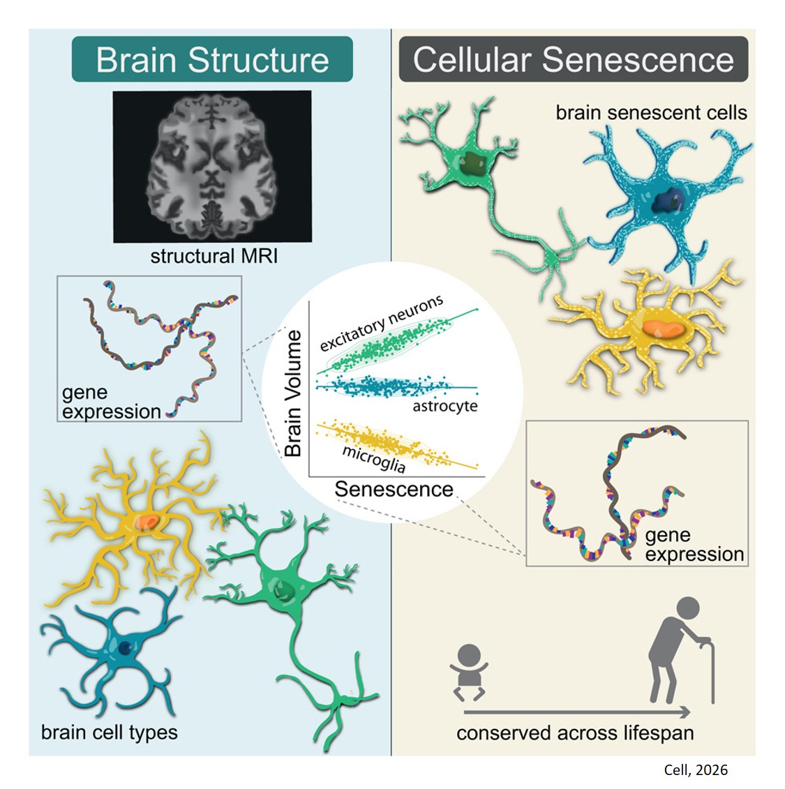
A key resource that was used as the discovery cohort in this study is the Living Brain Project, which uniquely pairs prefrontal cortex biopsies obtained during deep brain stimulation procedures with brain imaging data, allowing researchers to study molecular and structural features together in living individuals. The research team developed a method to define senescent cells in human brain tissue and used the resource to examine how senescence-related gene expression is associated with brain structure.
“This study addresses a major gap in the field by directly linking molecular features of the brain to neuroimaging measures in the same individuals,” said the other co-senior author of the paper. “By leveraging datasets from the Living Brain Project, we can begin to understand how senescence-related biology may differentially influence brain organization across cell types and across the lifespan.”
Among the study’s most striking findings was evidence that cellular senescence plays distinct roles in brain structure depending on cell type and stage of life. Genes associated with senescence in microglia—the primary immune cells in the brain—were linked to larger brain volumes, while senescence-related genes in excitatory neurons were associated with smaller brain volumes in the aging brain. Notably, the excitatory neuron findings were also observed early in life, providing the first evidence that senescence-related processes are at work soon after embryonic development.
"We were excited to see clear signs of senescence in both the aging and developing brain using our new method,” said the lead author of the study. “Our results support brain cellular senescence as an example of ‘antagonistic pleiotropy’—the idea that some genes help survival or fertility early in life but cause harm later, contributing to aging and disease. Most prior work links brain cellular senescence only to brain aging, but our finding of it during development shows this process is not just a marker of aging or disease; it also may play key roles in early brain development.”
“Often the greatest developments in medicine are not through invention of totally new means but through a unique understanding of whatever is already in reach,” said another author. “This work represents another fruition of the Living Brain Project’s ability to harness known data types in unique combinations to pave the way for future therapies. While brain ‘senescence’ or growing frail is largely accepted as a normal process of aging, this data set represents an opportunity to challenge that notion.”
While the findings do not point to immediate treatments, they offer a framework for understanding how brain structure changes over time and how age-related differences may emerge. The authors acknowledge the study’s limitations (relatively small and clinically specific cohort; focus on the prefrontal cortex; finding associations, not causal evidence) but are confident that their findings lay important groundwork for future research.
Future directions include expanding to larger and more diverse cohorts, refining cell type-specific definitions of senescence, and conducting experimental studies to determine whether senescence-related pathways causally influence brain structure. Together, these efforts could help clarify when and where senescence supports brain health—and when it may contribute to vulnerability in aging and in Alzheimer’s, Parkinson’s, and other neurodegenerative diseases.
https://www.cell.com/cell/fulltext/S0092-8674(25)01179-1
https://sciencemission.com/brain-cellular-senescence-and-brain-structure