

Mass spectrometry offers faster look at the edges of brain tumors
Glioma brain tumors are diffuse, so surgeons usually cannot excise all of the cancer during surgery. Currently, surgeons remove as much of the tumor as they can and then analyze the borders of the tumor after surgery. Researchers have developed a new method in which mass spectrometry could be used during surgery to evaluate tissue. This information could help guide the surgical team’s decisions, increasing the amount of cancerous tissue they are able to remove and improving the patient’s outcomes.
This research, which was recently published in PNAS.
Gliomas are highly infiltrative and deadly brain tumors with complex molecular characteristics that complicate diagnosis and treatment. Brain surgery is one of the most important treatments for glioma and can strongly affect a patient’s prognosis and survival. Because glioma cells can spread into nearby healthy brain tissue, particularly at the tumor margins, they are difficult to detect using conventional technologies available in the operating room. Tumor tissue that remains in the brain after surgery can contribute to recurrence and make further treatment more challenging.
“The information we are getting is currently not available under standard of care procedures,” said the lead author. “If our diagnostic method were to be approved, it would provide actionable molecular information during surgery. This information is currently not available on the time scale (some hours) of the surgery. This information could directly influence treatment, including the extent of tumor resection, although the final surgical decision would be made by the surgeon based on the overall clinical context rather than the mass spectrometry results alone. It could also help guide the selection of targeted therapies.”
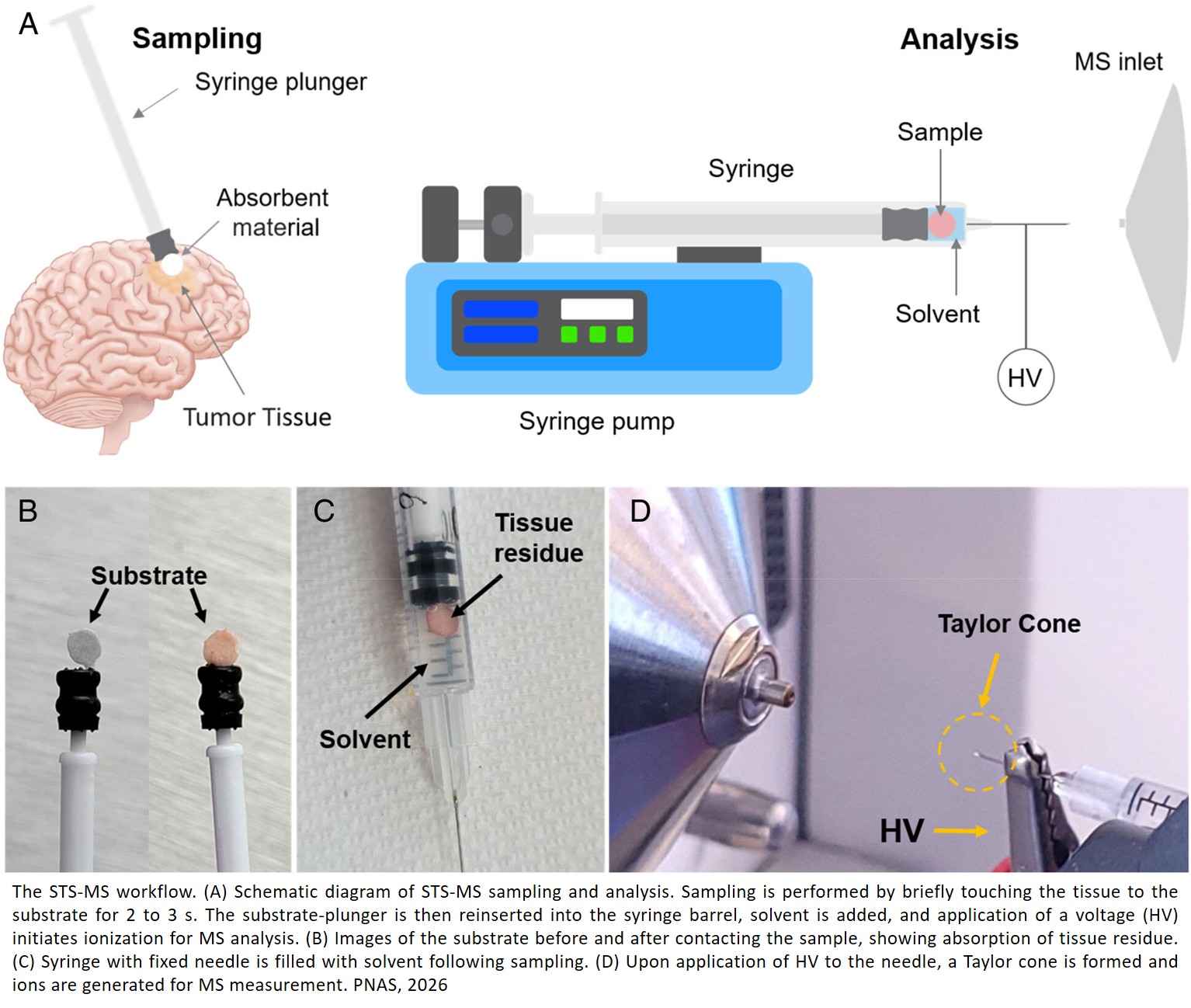
During brain tumor surgery, a small number of samples are taken from a surgeon-defined position in the brain for pathology. In this study, fractions of such samples were transferred onto the plunger of a hypodermic needle. Scientists would then spray a solution that measures the ratio of signals for multiple pairs of endogenous compounds in the brain using tandem mass spectrometry in a process that takes roughly three minutes.
“The study used 86 human brain samples, and the researchers showed that ratios of endogenous metabolites in brain tissue can distinguish glioma from non-tumor tissue with high confidence,” said the senior author. “It can also classify tumor subtypes and mutation status. The simple ambient ionization mass spectrometry approach enables fast, clinically compatible analysis with potential to improve margin assessment and enhance patient outcomes.”
Mass spectrometry is an analytical laboratory technique that measures the mass-to-charge ratio of ionized molecules to identify and quantify complex chemical compounds.
“The method was based on ambient ionization making ions from molecules in the open environment. This allows molecular measurements to be made in situ. The first such method was described in 2004 and has since been commercialized and widely used. The new method used in this study — syringe touch spray — uses an ordinary hypodermic to take a sample from the patient and perform mass spectrometry analysis.”
The samples analyzed in this study were taken using MRI-guided precision surgery. The tissue samples were characterized by standard methods, from which molecular information is available only after surgery.
Increased carnitine relative abundances were found across all glioma subtypes. Importantly, α-aminoadipate was significantly elevated in grade 4 glioma, which may correlate with increased tumor malignancy. Grade 3 gliomas showed markedly higher 2HG abundance than grade 4 samples. In addition, a selection of ratios showed a linear trend with varying tumor cell percentage.
Now that this has been tested on 86 human samples, the team will explore the next steps to get this technique into surgical situations and diagnoses. A similar 50-plus patient study will need to be undertaken in which the technique is used during surgery. According to the senior author, this would pave the way for a clinic trial to be proposed.
“In previous work, our measurements have been made in the hospital, during surgery (PNAS 2024),” said the author. “In this case, we recorded the mass spectrometry remotely and compared with the available clinical information. The very simple and quick measurement has high discriminating power, distinguishing diseased from non-diseased tissue as well as providing information on tumor grade and mutation status. This technology would not replace the current standard of care during brain tumor surgery. Instead, it could provide surgeons with molecular information that is not currently available in the operating room, helping to guide surgical decisions during tumor removal, particularly at the tumor margins.”
https://www.pnas.org/doi/10.1073/pnas.2616216123
https://sciencemission.com/Metabolite-ratiomics