

How loss of tumor suppressor p53 enables mutant cells proliferate through normal tissue?
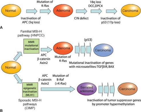
A protein known as p53 is, of all tumor suppressors in human cancer, the most frequently altered by gene mutations. When mutations cause it to be absent or nonfunctional, its loss can allow populations of mutant cells to expand through normal tissue. Researchers have long known that loss of p53 does more than remove a block to cell growth. But how p53 loss changes tissue behavior to permit this growth remains unclear.
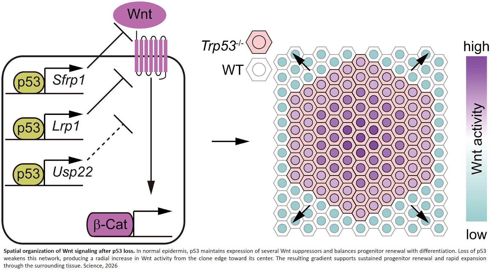
A research team found that loss of p53 can also change how growth and tissue development signals are organized across a population of mutant cells. In particular, loss of p53 reorganizes Wnt signaling, a major pathway that controls tissue growth and development, into a radial gradient, with lower activity near the edge of a mutant clone and higher activity toward its center.
Importantly, the researchers found that it is not simply the overall amount of Wnt signaling that determines how effectively mutant cells expand, but rather, how that signal is organized in space. Disrupting the Wnt signaling gradient sharply limits the ability of p53-deficient cells to overcome neighboring normal cells.
Using mouse skin as an experimental model, the researchers tracked how groups of p53-deficient epithelial cells, or clones, expand within otherwise normal tissue. By following these cells over time, they found that clonal expansion was driven primarily by a shift in cell fate: mutant progenitor cells were more likely to remain self-renewing and less likely to differentiate into mature cells. This effect, rather than simply faster cell division or reduced cell death, allowed mutant clones to continue expanding.
To uncover the mechanism behind this behavior, the team combined gene-expression analysis, mapping of where p53 binds to DNA, genetic screening of more than 1,000 candidate p53-regulated genes, and imaging of Wnt signaling at the single-cell level. They then experimentally changed the spatial pattern of Wnt activity to test whether clonal expansion depended primarily on how much Wnt signaling was present or on how that signaling was organized across the mutant cell population.
Loss of p53 shifted epidermal progenitor cells toward self-renewal and away from differentiation, allowing mutant clones to continue expanding through surrounding normal tissue. The researchers identified three genes directly regulated by p53—Sfrp1, Lrp1, and Usp22—that normally help restrain Wnt signaling.
When p53 was lost, Wnt activity increased and became organized into a persistent radial gradient. Clones with this gradient expanded efficiently, whereas clones with more uniformly elevated Wnt activity expanded less effectively, even when their overall Wnt activity was higher.
Most normal tissues accumulate cells carrying cancer-associated mutations as people age, yet many of these cells remain contained and never progress to cancer. This study provides a potential explanation for how loss of the major tumor suppressor, p53, can alter this balance and allow mutant cells to progressively colonize and take over normal tissue.
The findings also point to a broader principle in tissue biology: understanding how cell signaling pathways are arranged in space may be just as important as knowing whether those pathways are turned up or down. The work provides a framework for understanding how cancer-associated mutations can reshape tissue organization long before an overt tumor forms.
https://www.science.org/doi/10.1126/science.aed3065