


How cells turn mechanical forces into biochemical signals
Cells constantly probe their environments, searching for physical cues that guide their behavior. And yet a cell’s response to its environment is always biochemical, mediated by the chemistry of its internal protein machinery. So how does a cell convert mechanical information into a molecular process? It’s a long-standing mystery of cell biology, with various implications for cancer and other diseases.
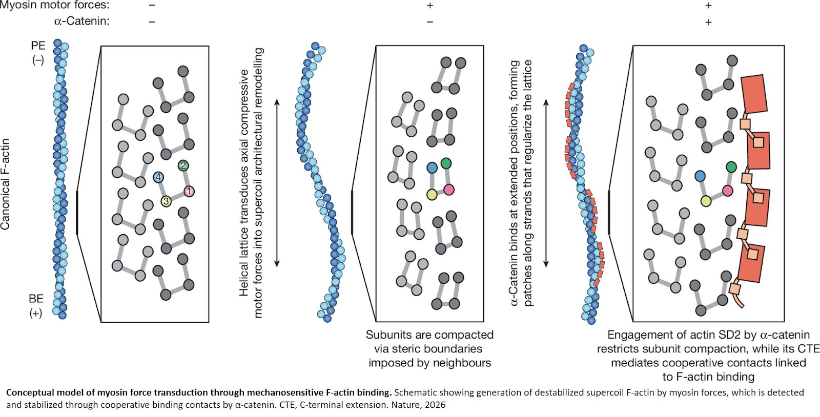
A research team has shown for the first time that when a motor protein called myosin compresses actin filaments within the cytoskeleton, it squishes the filaments into coils. This deformation is detected by protein sensors associated with cell adhesion, which congregate at specific sites on the cell interior. The findings were published in Nature.
“We knew that forces generated by myosin are critically important for cells to receive mechanical signals, but how that works hasn’t been clear,” says the senior author. “This is the first snapshot of a mechanical signaling complex in action.”
The cytoskeleton helps the cell transmit, receive, and process physical and biochemical information—a dynamic responsiveness that allows cells to interact with the world around them.
A key building material of the cytoskeleton is the actin filament, which powers cellular movement thanks to motor proteins like myosin, which tug, twist, and compress actin.
In 2020, the lab discovered that tugging on actin filaments with myosin actually helped the actin to bind better to a protein sensor, called alpha-catenin, which builds physical connections between cells. But they had no idea why.
Knowing why is important, because the better the binding, the better cells adhere to each other. “If you get rid of myosin, cells can’t stick together efficiently or transmit forces or information between them,” says the first author. “Everything just falls apart.”
The lab often relies on cryo-EM—a widely used imaging tool that takes snapshots at the resolution of a few angstroms, or billionths of a meter—but to observe myosin motor proteins exerting force on the filaments, they had to modify their usual approach.
They devised a way to secure myosin motors to the cryo-EM grid, fuel them up with ATP, and flash-freeze them as the myosin played tug-of-war with the actin filaments nearby. This approach allowed them to capitalize on a known characteristic of myosin motors: they fire randomly. That means they could capture a variety of states of action frozen simultaneously.
As a control, they removed the ATP fuel and repeated the process. “That way we could compare what happens in the presence and absence of fuel, and infer that the changes we see are due to the motor activity,” the senior author says. “That itself was a major technical innovation.”
Based on widespread assumptions in the field, they’d expected that tension imposed by myosin would be the key to priming the actin filaments for the alpha-catenin sensors. But, to their surprise, they instead found that compression was the key. This squeezing caused the filaments to turn into spirals—and it was this shape in particular that set off the alpha-catenin sensors.
And it was happening in a localized way. “Even if the entire network of myosin is generating tension—or tugging on the filaments—little segments of the network will actually be generating compression based on the random operation of the motors and how they happen to be positioned and firing asynchronously," the senior author describes. “That’s interesting, because it means these subpopulations could have a sort of signaling function.”
The authors also investigated how these coils might form using computer simulations. They ran simulations testing the three forces at play—tension, torsion, and compression—at various magnitudes and in different directions.
No matter the level of force or direction of action, they found the same result: compression was the key.
“This was also tricky,” the lead author says. “It wasn’t computationally difficult, but it required us to figure out how to capture the dynamics of a process that’s happening at an intermediate length scale— between the atomic level and the subcellular scale.”
While making fundamental insights into the processes that give our cells the ability to sense, move, and respond was a key result of the study—as were the technical advances that enabled those insights—the author notes that myosin dysfunction is connected to a number of diseases and that myosin inhibitors are in clinical trials for different conditions, including cancers such as glioblastoma.
“What studies like ours do is provide a way to potentially interpret which processes could be going wrong in certain diseases,” the senior author says. “At this level, causes are usually mysterious—for instance, why would having too much of a particular force on a protein be bad? Is it responding to these types of changes in the wrong way? Understanding the correct function of a process allows you to rationally design ways to correct dysfunctions in that process.”
https://www.nature.com/articles/s41586-026-10398-7
https://sciencemission.com/Myosin-forces-remodel-F-actin