


Cellular mechanism underlying microcephaly
Why do some children develop a brain that is too small (microcephaly)? An international research team has used human brain organoids to investigate how changes in important structural proteins in the cell lead to this severe developmental disorder (EMBO Reports).
Mutations in actin genes alter the way early progenitor cells divide in the brain. This reduces the number of these cells, which leads to reduced brain growth and smaller brain size. “Our findings provide the first cellular explanation for microcephaly in people with the rare Baraitser-Winter syndrome,” says the first author of the study.
Actin is a basic building block of the cytoskeleton, the internal support and transport structure of every cell. People with Baraitser-Winter syndrome carry a single gene mutation in one of two central actin genes. To investigate the effect of these mutations, the researchers generated induced pluripotent stem cells from skin cells of Baraitser-Winter syndrome patients. From these, they formed three-dimensional brain organoids that replicate important steps in early human brain development.
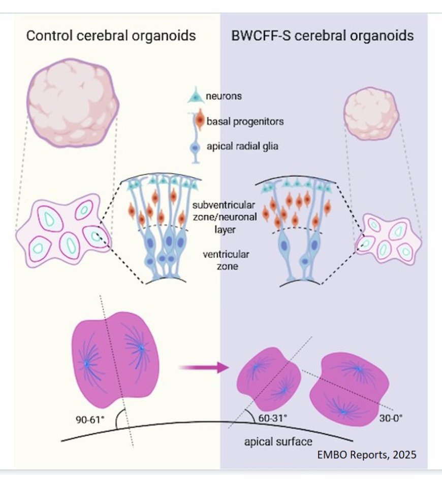
The results were clear: after thirty days of growth, the patient organoids were about a quarter smaller than the control organoids from healthy donors. The internal ventricle-like structures, where the progenitor cells are located and form early nerve cells, were also significantly smaller.
A closer look at the cell types in the organoids revealed a shift: the proportion of apical progenitor cells, i.e., the central progenitor cell population of the cerebral cortex, was significantly reduced. At the same time, there was an increase in basal progenitor cells, a type of daughter cell that normally only appears later in development.
The team used high-resolution microscopy to analyze the division of the apical progenitor cells. Normally, these cells divide predominantly perpendicular to the surface of the ventricular zone. Only in this way are the cell components distributed evenly and two new apical progenitor cells are produced. In the patient organoids, this very process was disrupted: the proportion of vertical divisions was massively reduced. Instead, the majority of cells divided horizontally or at oblique angles. This altered orientation meant that the apical progenitor cells renewed themselves less frequently, detached from the ventricular zone more often, and transformed into basal progenitor cells.
“Our analyses show very clearly that a change in the division orientation of the progenitor cells is the decisive trigger for the reduced brain size,” the last author of the study. “A single change in the cytoskeleton is sufficient to disrupt the course of early brain development.”
Electron microscope images revealed further abnormalities: the cell shapes at the ventricular surface were irregular. There were more protrusions between neighboring cells. In addition, there was an unusually high amount of tubulin, another component of the cytoskeleton that plays an important role in cell division, at the cell junctions. Although the basic cell architecture was still recognizable, these changes could be enough to permanently disrupt the division orientation.
To rule out the possibility that differences between patient and control organoids were caused by other genetic factors, the team conducted a control experiment: The healthy stem cell line was modified with CRISPR/Cas9 to carry exactly the same mutation as one of the Baraitser-Winter syndrome patients. The result: the brain organoids produced in this way showed the same malformations as the patient-derived organoids – a proof that the mutation itself is the cause.
“Our findings help us understand how rare genetic disorders lead to complex brain malformations and highlight the potential of brain organoids for biomedical research,” says the author.
"The therapeutic potential of this study lies in diagnostics, as our data helps to better classify genetic findings in patients. Since the disease affects early fetal development processes, interventions in humans would be complex. However, new drugs that influence the interaction between actin and microtubules could open up new approaches in the long term," says the Director of the Institute.
https://link.springer.com/article/10.1038/s44319-025-00647-7