

Regulation of protein transport from ER to Golgi
Cells synthesize proteins in the endoplasmic reticulum (ER), and these proteins are transported to the Golgi apparatus for distribution to their ultimate destinations inside and outside the cell. The structures that transport proteins from ER to Golgi apparatus are called COPII vesicles; the processes which regulate the formation of COPII vesicles are only partially understood.
An international research team has revealed how COPII vesicular transport between endoplasmic reticulum and Golgi apparatus is regulated during ER stress. This functions as a counterbalance to the negative effects of stress on protein synthesis.
Their findings were published in Nature Communications.
“We investigated how protein transport from the endoplasmic reticulum to the Golgi apparatus is regulated during endoplasmic reticulum stress,” says the corresponding author of the paper. “When cells are under stress, protein folding is disrupted, leading to the production of misfolded proteins. Consequently, cells possess a quality control mechanism that prevents these misfolded proteins from being released to locations other than the endoplasmic reticulum until they are properly repaired.”
Using the model organism Saccharomyces cerevisiae, budding yeast, the researchers created a series of mutants for genes associated with COPII vesicle-mediated transport and analyzed their phenotypes.
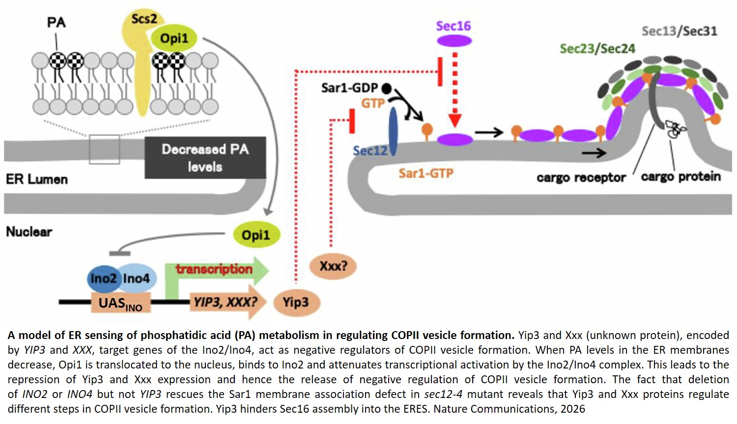
The researchers found that endoplasmic reticulum stress leads to an increase in the levels of phosphatidic acid (PA) in the endoplasmic reticulum membrane. Furthermore, they demonstrated that this change in PA levels increases YIP3 gene expression via the Opi1–Ino2/Ino4 transcriptional regulatory system, and that the Yip3 protein limits COPII vesicle formation by suppressing the accumulation of Sec16 at the ER exit site (ERES), the site of COPII vesicle formation.
“The key point we wish to convey is that when cells experience endoplasmic reticulum (ER) stress, they possess a mechanism that detects changes in their own membrane lipid state and suppresses intracellular transport via transcription,” says another author. “In this study, we demonstrated a mechanism by which the endoplasmic reticulum (ER) uses changes in the membrane lipid PA as a cue to increase Yip3 levels via transcription, thereby suppressing the accumulation of Sec16—a scaffold protein for COPII vesicle formation—at ERES, and consequently inhibiting COPII vesicle-mediated transport.
This discovery indicates that the ER possesses a sophisticated mechanism for regulating intracellular trafficking in response to stress.” Scaffold proteins are proteins that tether multiple proteins to a specific cellular location, thereby enhancing the efficiency of reactions mediated by protein complexes.
Under normal conditions, Sec16 accumulates on the ER membrane, where it functions as a platform for the “shipping gate” for intracellular transport; under endoplasmic reticulum (ER) stress, Sec16 no longer accumulates at ER exit sites (ERES). The finding that changes in membrane lipids regulate the assembly of transport machinery via transcription represents a novel concept that had not been previously reported.
“The next step is to identify which molecules Yip3 uses to suppress the accumulation of Sec16 at ERES,” the author concludes. “Ultimately, we aim to elucidate how the ER senses stress and membrane lipid states and how it transmits this information to each stage of COPII vesicle formation, with the goal of fully understanding the regulatory mechanisms of intracellular transport during ER stress.”
https://www.nature.com/articles/s41467-026-75057-x
https://sciencemission.com/ER-sensing-of-lipid-metabolism