


Developing tumor microenvironment for cancer growth
Scientists are shedding new light on a tumor’s earliest moments — revealing how lung cells with cancer-causing mutations recruit accomplices from healthy surrounding tissue to pave the way for a tumor to develop.
This corruption of the local neighborhood — what scientists call the “tumor microenvironment” — begins surprisingly early, as tumors first emerge, according to a study published in Nature.
The team’s findings show that when this communication with surrounding cells is disrupted, tumors fail to grow.
“We also found that this transformation of the local neighborhood is reversible, if caught early enough. This opens the door to new treatment and prevention strategies,” says the study senior author.
The research was conducted in mouse models of lung cancer carrying KRAS mutations — one of the most common genetic changes in the disease — as well as in 3D “assembloids,” miniature organs created from mouse and human lung tissue.
The research builds on previous work studying how healthy lung tissue responds to injury.
Normally, when a lung tissue is damaged, specialized stem cells enter a regenerative state where they become flexible so they can replace the damaged cells. Then, once the injury is repaired, they go back to normal.
But when these stem cells acquire a cancer-causing mutation in the KRAS gene, they get stuck in this regenerative state and multiply out of control, creating tumors.
Using sophisticated experiments that allowed them to track individual tumor cells from the moment they acquired KRAS mutations, the team was able to map what happens next with remarkable precision.
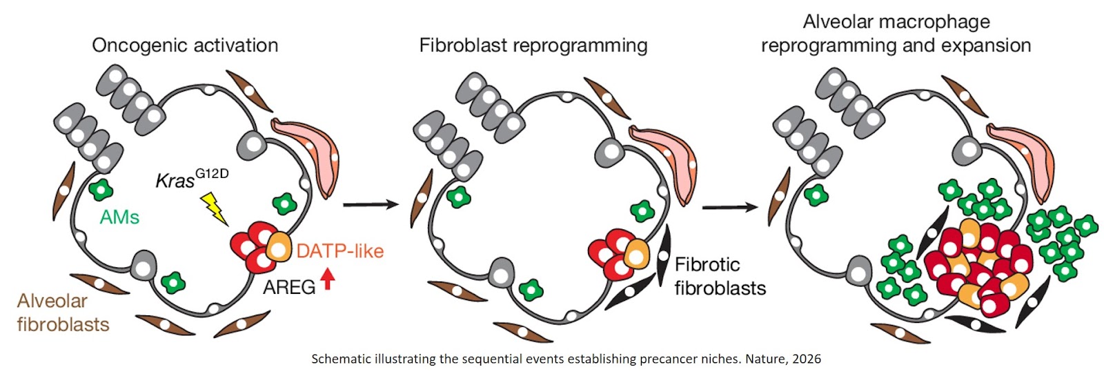
What they discovered is a three-step process:
· First, the mutant cells enter regenerative states and produce high levels of a protein called amphiregulin (AREG). “This is a distress signal, which is received by nearby healthy cells,” the senior author says.
· Second, connective tissue cells called fibroblasts receive this signal and act as if they are responding to an injury. The fibroblasts become “fibrotic,” producing a fibrous scaffold around the tumors, as they would during normal wound healing.
· Third, the activated fibroblasts help tumor cells grow and also send their own signals that reshape the local immune response. Macrophages expand at the site and actually suppress the immune response, rather than attacking the mutant cells. It happens because these reprogrammed cells call in the wrong backup — instead of attracting cancer-fighting immune cells, they recruit cells including neutrophils and regulatory T cells, whose normal role is to quiet things down and prevent the immune system from overreacting and damaging the body’s own tissues.
The result is a self-reinforcing loop, the lead author says. The remodeled environment helps tumor cells maintain their regenerative state, while the tumor cells continue to send out their distress call, which reshapes the tissue around them. Together, these conditions create a conducive environment for a tumor to grow and progress.
The researchers then asked a crucial question: What happens if you disrupt this communication network between cancer-causing cells and the normal cells in their neighborhood?
They used an EGFR inhibitor approved for treating advanced lung cancer with EGFR mutations to block the AREG distress signaling — and the results were dramatic. The fibroblasts remained normal, the undesirable immune response didn’t occur, and tumor development was severely impaired. The team also saw similar results when they deleted the AREG gene from mutant cells. Without the distress signal, fibroblasts and immune cells remained in their normal states, and tumors failed to develop.
Even more remarkably, when they blocked KRAS activity in early lesions that had already formed, many of these changes reversed.
“Beyond genetic alterations, communication with nearby healthy cells to build a ‘pre-cancer niche’ is essential for tumors to grow,” the senior author says. “Encouragingly, at these early stages, those altered surrounding cells can still switch back to normal.”
Findings in laboratory models, however, don’t always hold true in actual patients. So the team took steps to test the applicability of their findings.
When they analyzed tissue samples from patients with early-stage lung adenocarcinoma, they found the same key players positioned close together: cancer cells producing high levels of AREG and adjacent fibrotic fibroblasts.
But patient tissue samples can only provide a single snapshot in time — they can’t show the sequence of events as cancer develops.
To overcome this, the team developed an innovative assembloid system. They isolated healthy lung stem cells from patients’ lungs and grew them into 3D miniature organs in the laboratory. By introducing KRAS mutations into these cells, they were able to watch the earliest steps of tumor development in real time.
“For the first time, we can trace how healthy human lung cells begin to develop cancer when they acquire cancer-causing mutations,” the senior author says. “KRAS mutation drives these cells into the same AREG-high regenerative state we observed in the mouse model.”
When the team co-cultured these KRAS-mutant organoids with normal human lung fibroblasts, they saw the same chain of events: fibroblasts became fibrotic, and this transformation could be blocked with the same EGFR inhibitor.
Additionally, the team wanted to understand whether this behavior was specific to KRAS mutations or represented a more general mechanism.
They ran the same tests using another common lung cancer mutation in the EGFR gene. And once again they found the same pattern.
Other research groups have also found similar results in esophageal cancer and pancreatic cancer, the author notes, strongly suggesting a broader pattern of tumor cells quickly building a protective environment that helps sustain their growth.
Although the clinical implications of the work are still at an early stage, the findings point to potential biomarkers that could help detect lung cancer much earlier. If validated, these markers could enable diagnosis at stages when the disease is far more treatable, the author adds.
They might be especially relevant for people at high risk of developing lung cancer — such as long-term smokers or those with genetic predispositions — who might unknowingly harbor precancerous cells with oncogenic mutations.
“There are clinical reports that some patients with high-AREG tumors respond to EGFR inhibitors, even when their EGFR genes don’t harbor mutations,” the author says. “Our work may help explain these cases and suggest new opportunities to identify and treat patients earlier.”
Overall, the work exemplifies an important shift in how we think about cancer’s origins, the author says.
“The key message we’d really like to deliver is: When tumors first emerge, they hijack the regenerative program, and they interact with healthy cells to generate a microenvironment supporting the cancer,” the author says. “This determines whether these tumor cells sustain and progress — and it happens at a very, very early stage.”
Rather than viewing cancer solely as a disease of mutated cells growing out of control, this research is part of an emerging understanding of cancer as a disease of corrupted cellular communication — one where mutant cells manipulate their healthy neighbors into providing support.
https://www.nature.com/articles/s41586-026-10399-6
https://sciencemission.com/tumour-permissive-microenvironment