

Where and how Alzheimer's tangles begin
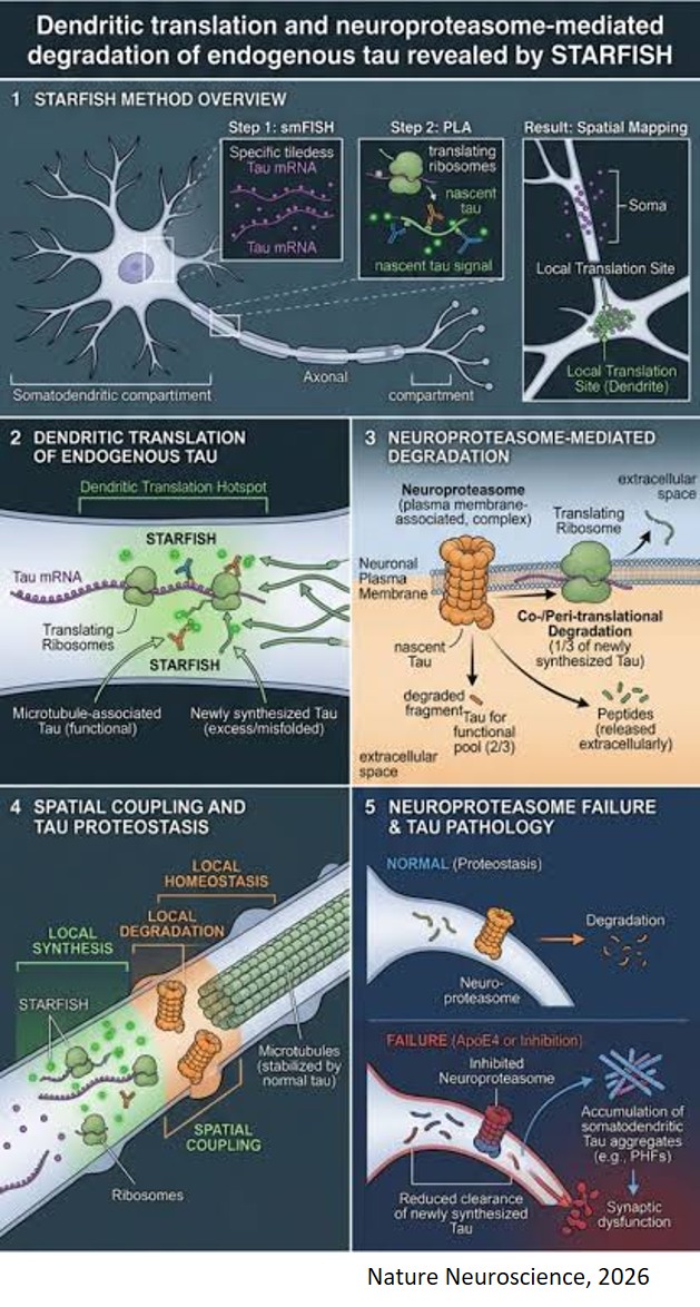
Where and how does an Alzheimer’s tangle begin? A new study by neuroscientists challenges a long-held assumption about the source of toxic tau proteins, the protein that forms neurofibrillary tangles, that are closely linked to memory decline in Alzheimer’s disease.
The study reveals a previously hidden first chapter in the life of tau: its unexpected birthplace in the dendrites, the branching extensions that neurons use to receive signals from other cells. The study also identifies a rapid quality-control process that destroys a substantial fraction of new tau and may help explain how normal tau takes a pathological turn.
These findings run counter to decades-old assumptions about the first steps of tau pathology in Alzheimer’s disease.
“If we want to stop tau pathology before tangles form, we need to understand the first moments of tau's life,” says the study’s senior author. “We found that tau is born in dendrites and placed under intense quality control almost immediately. That gives us a new place and a new moment to look for how pathology begins.”
In healthy neurons, tau proteins are normally concentrated in axons, the long projections that transmit signals. In Alzheimer’s disease, tau accumulates in the cell body and dendrites and clumps together into filaments and tangles. The prevailing explanation has been that mature axonal tau detaches from microtubules and redistributes into these compartments. Many researchers are trying to find ways to block the travel of tau to prevent or treat the disease.
But the new study, published in Nature Neuroscience, proposes a very different explanation for tau’s presence in the dendrites: The proteins are born there.
Using a new imaging technique the researchers developed to pinpoint the birthplace of any protein, the team found that tau proteins are synthesized solely in a neuron’s dendrites.
Almost immediately, the newly created tau is subject to intense quality control, and within minutes, a third of new tau proteins are disposed of by the neuroproteasome, unusual protein-disposal machines located at the neuronal plasma membrane.
“It gives us a completely different picture of the cell biology of tau proteins and how they transform into aggregates,“ the author says. “The field has largely focused on tau moving to the wrong place. Instead, we found that a vulnerable pool of tau is being made in dendrites all along. The question becomes what normally keeps that pool safe, and what changes when that protection fails.”
Instead of harmful tau proteins traveling from axons to dendrites, the authors propose that a harmful form of tau is created locally in the dendrites when newly synthesized tau is not folded properly. The researchers found neuroproteasomes positioned beside many of the ribosomes translating tau, as if neurons had installed quality control next to the assembly line.
This hypothesis, which builds on lab's earlier work on the neuroproteosome’s role in tau processing, suggests that misfolded tau proteins are usually eliminated through the neuroproteosome. But when the quality-control machinery breaks down, misfolded tau escapes disposal and begins assembling into the characteristic filaments seen in Alzheimer's disease.
“I think what we’re showing here is that there are mechanisms that control tau synthesis and folding that are relevant to Alzheimer’s disease,” says the author. “If we can understand these pathways and triage systems, we may find new ways to ensure that tau folds properly and prevent it from taking a pathological turn.”