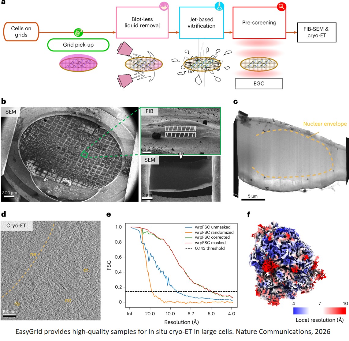


Mechanism of Blood-Brain Barrier disruption by Extracellular Vesicles in Preeclampsia
Cerebrovascular complications in preeclampsia is unknown.
The authors investigated if circulating small extracellular vesicles (sEVs) generated from preeclampsia (PE) or placentae cultured under hypoxic (Hyp) conditions impaired the expression of tight junction proteins, such as CLDN5 (claudin-5) mediated by VEGF (vascular endothelial growth factor), and activation of KDR (VEGFR2 [VEGF receptor 2].
The authors show that sEVs-PE disrupts the blood-brain barrier, an effect replicated with sEVs-Hyp, and involves reduced CLDN5 and elevated VEGF contained within these vesicles.
They also demonstrate that KDR activation did not result in the downregulation of CLDN5 observed with sEVs-Hyp.
These findings will improve our understanding of the pathophysiology of cerebrovascular alterations in women with preeclampsia.
https://www.ahajournals.org/doi/full/10.1161/ATVBAHA.124.321077