


Cell-type specific TDP-43 pathology in the motor cortex
Amyotrophic lateral sclerosis (ALS) and frontotemporal dementia (FTD) belong to a spectrum of neurodegenerative diseases with overlapping symptoms, characterized by muscle wasting, paralysis, dementia, and other serious impairments. There are currently no effective treatments. Many patients have a common hallmark: A protein called TDP-43 clumps together in the neurons of the brain to form tiny lumps.
Researchers have now discovered that these pathological changes primarily affect certain cells. Their findings, published in the scientific journal Nature Communications, could contribute to the development of new therapies.
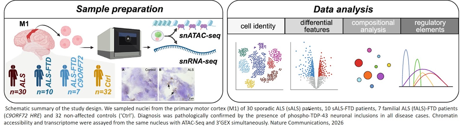
For their study, the research team examined brain tissue from deceased patients with ALS, with a mixed form of ALS and FTD, as well as from individuals who had not shown neurological symptoms during their lifetime. All samples came from the “motor cortex”, a brain area responsible for movement control. In total, neurons from around 80 individuals in Germany, the Netherlands, Scotland, and the United States were analyzed using advanced techniques.
“In ALS, as well as in the mixed form of ALS and FTD, TDP-43 deposits occur in different regions of the brain. However, the motor cortex is particularly relevant for movement disorders, which is why we focused on this area,” explains the research group leader.
The researchers found that not all neurons in this area are equally affected. “The protein aggregates occur predominantly within excitatory cells, that is, within neurons that serve to transmit and amplify nerve signals. These cells seem to be particularly susceptible to the disease. This phenomenon is referred to as selective vulnerability and known for a long time in the field. In addition, within the affected neurons, we found five subgroups (intratelencephalic L2-L3-LINC00507-FREM3, L3-L5-RORB-LNX2, L3-L5-RORB-ADGRL4 & L6-THEMIS-LINC00343 neurons and extratelencephalic L5-FEZF2-NTNG1 neurons). Each of these is impacted by the disease in a specific way,” explains the author.
The findings are based on the “transcriptome” of affected neurons. This molecular fingerprint provides information about which genes are active in affected cells and therefore enable to distinguish pathological processes in different cells. Based on this signature, the researchers were also able to identify cell type-specific changes. “Our data offer insights into disease mechanisms and thus point to possible targets for therapy development. For example, one can see how the activity of certain genes is altered depending on the cell type. The observation that not all neurons are equally affected suggests that future therapies will need to be tailored to specific cell types in order to combat the disease effectively,” says the author.
https://www.nature.com/articles/s41467-026-69944-6
https://sciencemission.com/TDP-43-pathology