


Imaging immune cells for human kidney disease
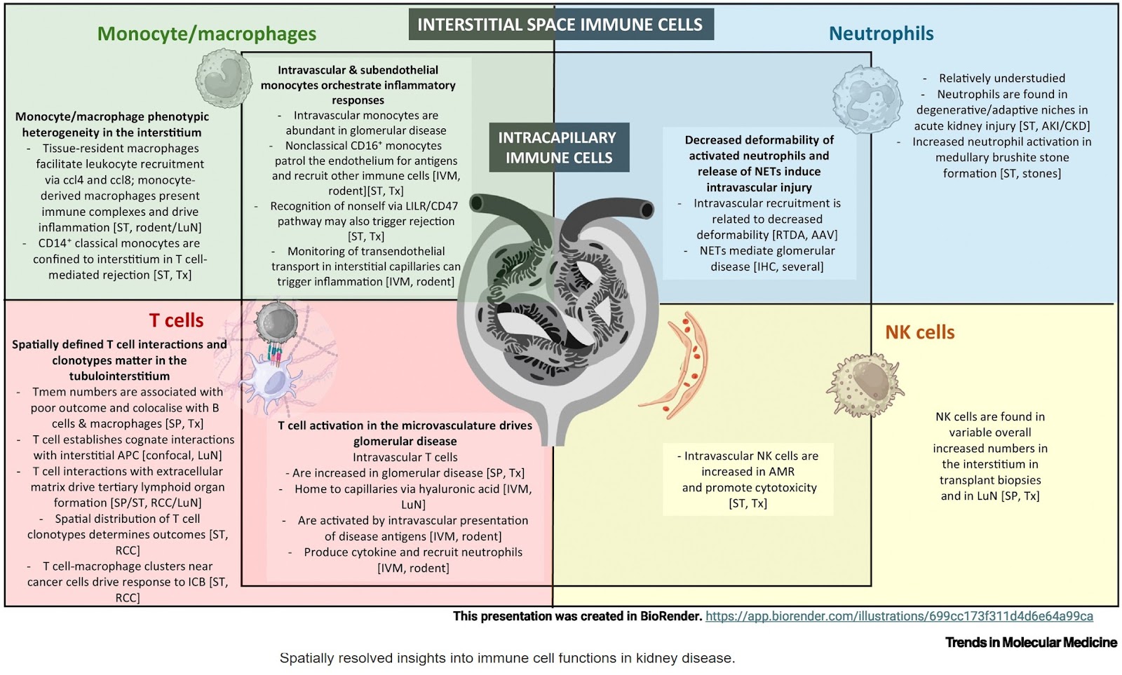
Cells of the adaptive and innate immune systems initiate kidney disease within specific niches, such as within tumors, within glomeruli (glomerulonephritis), or within the microcirculation (kidney transplants).
Novel advanced imaging techniques can investigate these niches using the ability to image immune cell behaviors in vivo (intravital microscopy) or ex vivo (e.g., neutrophil deformability assays); spatially resolved multiplex protein or transcript expression (spatial proteomics or transcriptomics); or beyond usual light microscopy (e.g., X-ray, confocal, light sheet, etc.).
They have revealed compartment specific immune architectures relating to monocyte trafficking and antigen surveillance; T cell trafficking, clonality, and cognate interactions; heterogeneity of cell subtype distribution across compartments; and neutrophil NETosis.
Cross-scale multimodal imaging pipelines are being designed to investigate these niches, combining lower resolution imaging with ultrastructural and molecular detail.
https://www.cell.com/trends/molecular-medicine/fulltext/S1471-4914(26)00091-2