










Neck lymph node removal linked to increased risk of cognitive decline
Surgery to remove lymph nodes in the neck may interfere with cerebrospinal fluid (CSF) drainage from the brain, contributing to cognitive impairment, according to a new multicenter research.
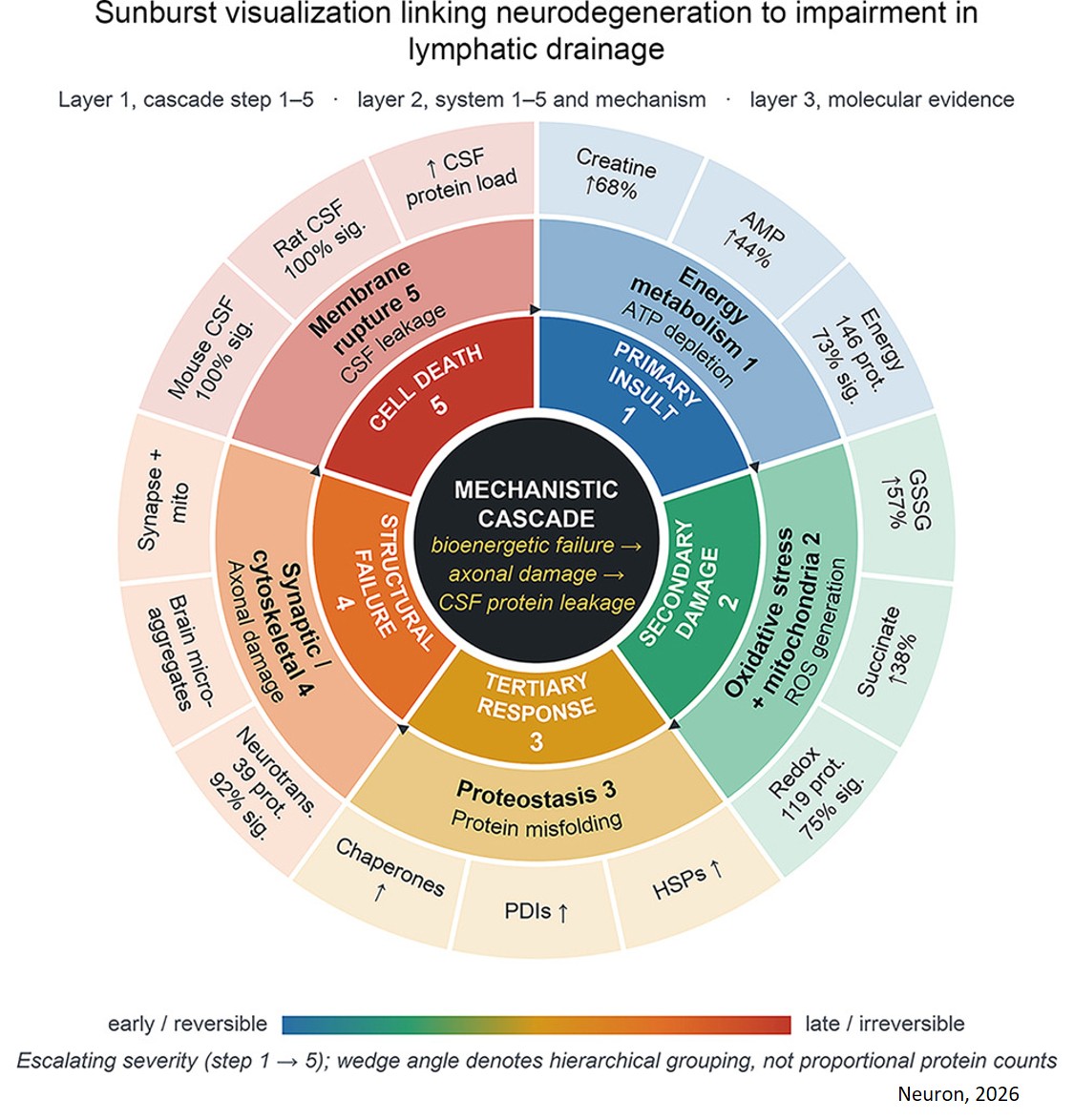
The paper, published in Neuron, found that patients with head and neck cancer undergoing cervical lymphadenectomy, a procedure to remove nearby lymph nodes to reduce cancer spread, had a higher rate of cognitive impairment than the general population. The researchers went on to demonstrate in preclinical studies how the procedure can disrupt the brain's waste-clearance system, leading to fluid buildup and a molecular process called oxidative stress that damages cell proteins and triggers inflammation.
"Your body produces lymphatic fluid to wash away waste from every organ, including the brain," said the senior author. "If this system is lacking, negative consequences may eventually build up.”
Neck lymphedema, or swelling caused by disrupted lymph drainage, is a common complication in patients after lymphadenectomy, said the senior author. However, this is one of the first papers to fully address what happens when lymphatic fluid cannot be properly cleared from the brain.
The researchers analyzed records of 1,035 patients who underwent neck lymphadenectomy without additional chemotherapy or radiotherapy at Montefiore Einstein. Rates of cognitive impairment were higher than estimates reported for age-matched populations.
About 18% of patients younger than 65 experienced cognitive impairment within two years after the procedure. In the general population, the overall risk of developing mild-to-severe cognitive impairment in this age group is 4 to 6%.
For patients older than 65, 25% of the surgery group developed cognitive impairment, compared to 10 to 22% of the general population. While the findings suggest an association between cervical lymphadenectomy and later cognitive impairment, further research is necessary to confirm a direct link.
The researchers also studied a second group of 59 patients who underwent cervical lymphadenectomy with no radiation or chemotherapy.
These patients had brain MRIs both before and after their surgeries. The imaging showed accelerated widening of a structure called the ventricular temporal horns after surgery, indicating brain atrophy or shrinkage. This was pronounced when lymph nodes were removed from both sides of the neck.
To uncover the biological mechanisms that might contribute to the observed cognitive decline, researchers conducted studies in animal models. When fluid could no longer drain normally from the brain, researchers found evidence that brain cells were under increased oxidative stress and that proteins associated with inflammation increased.
Interestingly, the harmful molecular changes appeared after sudden disruption of lymphatic drainage and were not detected when drainage declined gradually in a separate animal model.
They also found that proteins like synapsin, which are involved in relaying signals between neurons, were damaged and clumped together. These dense clumps differed from the plaques of amyloid-beta protein and thin threads of tau protein typically seen in Alzheimer's disease.
This retrospective study evaluated records of patients who already had treatment. Next, the researchers plan to conduct a prospective study, which follows patients over time as they undergo cervical lymph node removal, the author said.
Surgeons are already exploring ways to improve lymphatic drainage that may one day be applied to those with head and neck cancer at risk for cognitive problems. “Research into transplanting a lymph node from another area of the body may offer a way for lymph fluid that is blocked to enter the venous system and prevent lymphedema,” the author said.
https://www.cell.com/neuron/fulltext/S0896-6273(26)00688-4
https://sciencemission.com/Cervical-lymphadenectomy