


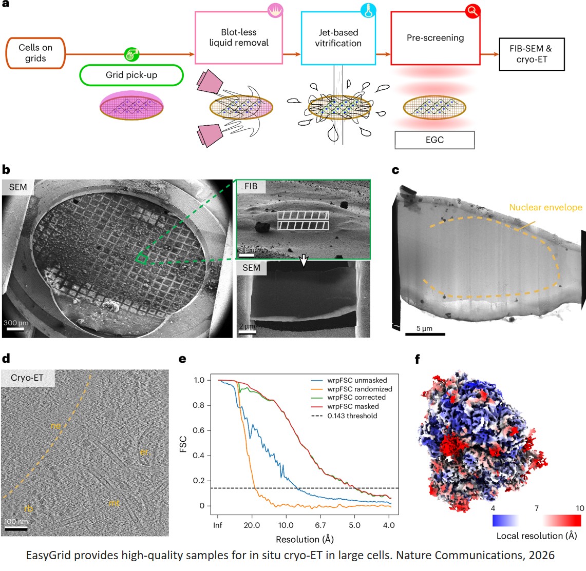


Platform for automated cryo-EM sample preparation and quality control
Cryo-electron microscopy (cryo-EM) can help scientists determine the three-dimensional structure of proteins in unprecedented detail.
Despite recent advances in cryo-EM methods, preparing high-quality samples remains challenging. The team, which specialises in creating innovative instrumentation for structural biology, is currently addressing this problem.
The team developed two unique systems, EasyGrid and EasyGrid Control, which they describe in a new Nature Methods publication. These platforms automate the preparation of high-quality samples for studies across scales – from single particles to whole cells – and can be adapted to different techniques: cryo-EM, cryo-electron tomography (cryo-ET), and X-ray nano-imaging.
The primary limitation to preparing high-quality vitrified samples is the manual preparation of samples, which requires careful handling and suffers from a lack of reproducibility in ice thickness and ice quality. If the ice layer is insufficiently thin or full of ice crystals, the electron beam cannot penetrate it or deviates due to diffraction, rendering the sample unsuitable for further imaging.
In addition, there is no way to easily pre-screen and check the quality of vitrified samples before viewing them using a cryo-electron microscope. These limitations make for poorly reproducible, lengthy, and often costly experiments.
The team, which has previously automated processes in X-ray crystallography – now used in most synchrotrons worldwide – took on the new challenge of improving cryo-EM sample preparation processes in 2017.
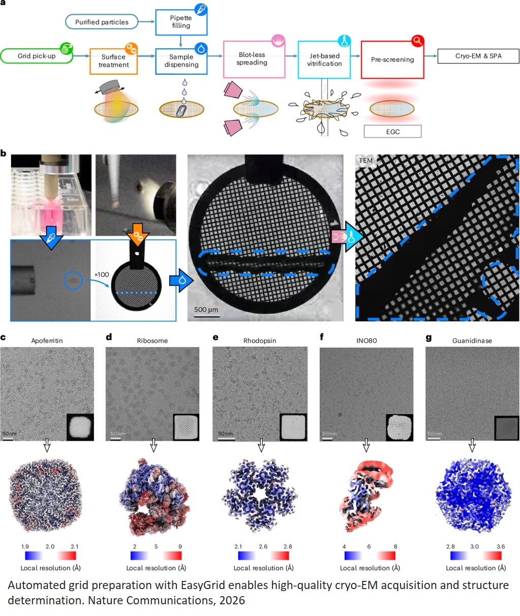
After several years of iterations on a first prototype, the team created EasyGrid – a fully automated and modular platform for high-throughput sample vitrification. In parallel, they developed EasyGrid Control – a unique instrument capable of automatically screening samples prepared with either EasyGrid or traditional methods to check their quality before use in advanced microscopy.
“With EasyGrid and EasyGrid Control, the whole process line, from the loading of the grid to the screening, is done autonomously,” explained a co-first author of the publication. “This leads to a more repeatable process and allows us to optimise samples faster and measure their quality, so we only load the best grids into the electron microscope”.
Another important aspect of EasyGrid is the use of different sample dispensing and vitrification methods from those currently offered by other sample preparation techniques.
The system uses pressure waves to spread the sample across the grid’s surface, enabling reliable liquid-layer thinning for single-particle analysis. It can also do this for samples comprising cells cultured directly on the surface of the sample holder grid or in suspension, dispensed with automated pipettes.
Additionally, while conventional preparation methods usually involve immersing the grid in liquid ethane, the team has developed an ethane jet-based vitrification system using bare grids (grids that are not mounted in an autogrid cartridge, a kind of circular copper frame). "This dramatically improves the cooling rate, enabling vitrification deeper within the cell, including the nucleus,” said the author. Hence, this sample preparation method is also advantageous for in situ (in the context of the cell) studies using other imaging technologies, such as cryo-electron tomography and X-ray nano-imaging.
Throughout the project, the team obtained high-resolution maps of several macromolecular complexes, including apoferritin, yeast ribosomes, guanidinase, the INO80–nucleosome complex, and pentamers of the bacterial rhodopsin KR2. The team also demonstrated a sevenfold improvement in the vitrification quality achieved within the nuclei of large human cells, compared with traditional plunge-freezing methods.
“Such a platform is an important game-changer not only in the quest for reliable automated cryo-preparation for high-throughput analysis by cryo-EM, but also for other types of atomic-resolution imaging of biological samples preserved in their native hydrated environment,” said a senior researcher.
“We evaluated the platform within our project on intracellular metal imaging, focusing both on the fate of theranostic nanoparticles [small particles that can be used simultaneously for medical diagnostics and therapy] and on how metal ions influence protein aggregation,” added the author. “The method delivered robust, reproducible cryo preparation, embedding cells in a very thin, homogeneous layer of amorphous ice. It also adapts seamlessly to our custom sample supports – square silicon nitride windows used for synchrotron cryo X-ray fluorescence nano imaging – resulting in superior image quality.”