


Mapping the lipid composition of ER membranes
Human cells contain billions of molecules. To prevent complete chaos, these molecules and the reactions between them are organized within structures known as organelles. One of the cell’s most important organelles is the endoplasmic reticulum, a dynamic network that extends throughout the cell and plays an essential role in cell growth.
Using a method they developed themselves, an international team of molecular biologists and bioinformaticians has tackled the challenge of determining precisely which molecules make up the membrane of this organelle. The researchers have published their findings in the highly regarded journal EMBO Reports.
‘Metabolic disorders occur when biochemical processes in the body are disrupted, thereby disturbing the conversion and distribution of nutrients. A wide range of complex metabolic disorders originate in the endoplasmic reticulum, where the cell produces most membrane proteins and synthesizes membrane lipids. To develop effective, targeted therapies, we need to understand how the endoplasmic reticulum responds to metabolic imbalances,’ says the corresponding author of the study. ‘The endoplasmic reticulum is of particular interest because it acts as a highly sensitive metabolic “sensor”, responding both to cancer and to chronic inflammatory diseases such as rheumatoid arthritis and Crohn’s disease,’ explains the author.
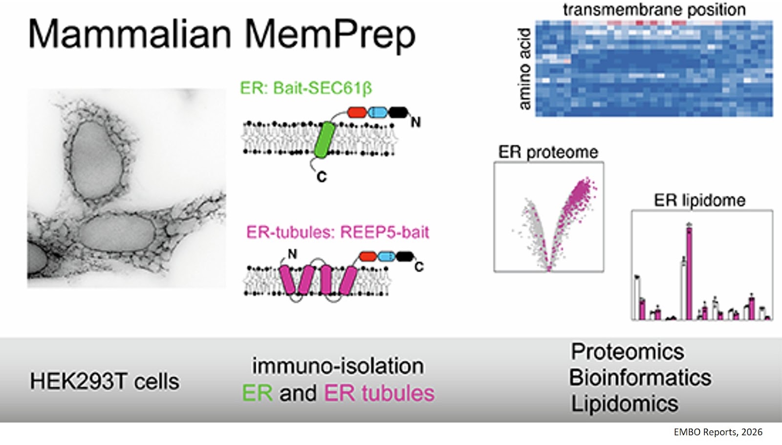
The research team has therefore developed a method that enables the endoplasmic reticulum to be isolated from the cell and then profiled using state-of-the-art analytical techniques. ‘The biggest challenge was that the endoplasmic reticulum is physically connected to virtually every other organelle in the cell to facilitate rapid organelle communication. To obtain pure preparations, we first had to break these connections before we could use antibodies to extract the endoplasmic reticulum,’ explains the first author.
By refining their MemPrep technology, which was originally developed for the model organism yeast, the researchers have now succeeded in isolating and characterizing the organelle from mammalian cells. This enables them to investigate in detail how the endoplasmic reticulum responds to metabolic stress and identify potential ways of restoring metabolic balance in stressed cells.
The study is also of particular interest to cell biologists, as it provides evidence that different regions of the endoplasmic reticulum are composed of the same membrane lipids regardless of whether they form tubules or sheets. ‘This was a surprising result, as these different regions are clearly stabilized by different proteins, yet their membrane-lipid composition remains unchanged,’ says the senior author.
They also demonstrate that the physicochemical features of ER lipids mirror those of ER-resident membrane proteins, including reduced hydrophobicity and increased polarity compared to plasma membrane proteins.
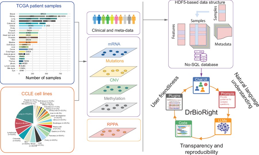
‘Mapping the cell and its organelles is an important step towards creating a digital twin that will enable us to simulate cellular processes on a computer and generate predictions that can then be tested experimentally in the laboratory. The results of these experiments can, in turn, be used to refine the digital twin in a continuous iterative process,’ explains the senior author.
https://link.springer.com/article/10.1038/s44319-026-00952-9
https://sciencemission.com/Mammalian-MemPrep