




How a protein complex guards the entrance to channels in mitochondrial membrane cristae
Twenty-four hours a day, seven days a week, mitochondria in our cells churn out energy from the food we eat, which keeps our bodies functioning. Much of this work happens along cristae — deep pockets in the mitochondria’s inner membrane. To work properly, cristae must maintain their shape, while carefully controlling which molecules enter and exit. At their narrow entrances sits a protein complex called MICOS, which helps stabilize these pockets and acts as a molecular filter. When this architecture is disrupted, the consequences can include neurodegenerative diseases and cancer. But exactly how MICOS works has remained unclear.
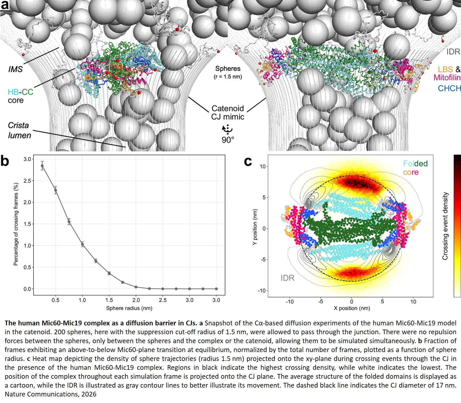
A research team has now modeled the human Mic60-Mic19 subcomplex, a central part of MICOS. Their simulations, which were published in “Nature Communications,” show how its flexible structure spans the cristae entrance, allowing smaller molecules to pass through while blocking larger proteins.
“Our study not only reveals the molecular architecture of an essential cellular machine, but it also shows how one can model highly dynamic protein complexes that escape traditional structural biology methods,” says a co-senior author of the study.
MICOS is made up of multiple copies of several proteins. Its largest component, the Mic60-Mic19 subcomplex contains a long, disordered region — a flexible stretch with no fixed shape. This flexibility is one reason it has been difficult to image or model.
To build their model, the team combined several approaches. They first captured the structure of a section of Mic60 found only in animals using X-ray crystallography. They combined this structure with fungal structures and AI predictions to build a virtual model of the human version. The model, however, had a big weakness: it was static, so it did not show the subcomplex’s flexibility. The team then simulated computationally the Mic60-Mic19 subcomplex’s movements.
To test how well their model replicated the actual human subcomplex, they compared it to structural data from human mitochondria. They found 97% correspondence, suggesting that their model closely reflects the structure of the actual human subcomplex.
Finally, the team tested the subcomplex’s gatekeeping function by adding spheres to simulate the passing of different sized proteins. They found that the subcomplex blocked spheres with a radius larger than two nanometers. Its disordered regions were key to this function: “With one static structure, we might not have seen exactly how it swats all of those spheres away,” says the author. “That's exactly why we needed an approach to see it in action.”
“Computational methods often produce impressive images and videos of molecular machines in action, but they are difficult to prove experimentally,” adds the author. “Our study now provides a way to do this.”
Mitochondria are essential in all cells, but tissues that need a lot of energy, such as muscle and the brain, are especially affected when they fail. The researchers found that a known mutation in patients with optic nerve damage and a developmental brain disorder alters the MICOS complex's core. Their model offers a possible explanation for how the mutation causes disease.
Confirming this will require observing the entire complex at work inside mitochondria. So far, the team has modeled only part of it. “We are now looking into the structure of the machinery inside cells, and how it changes during disease and aging,” says the author.