

How myeloid cells enter the central nervous system
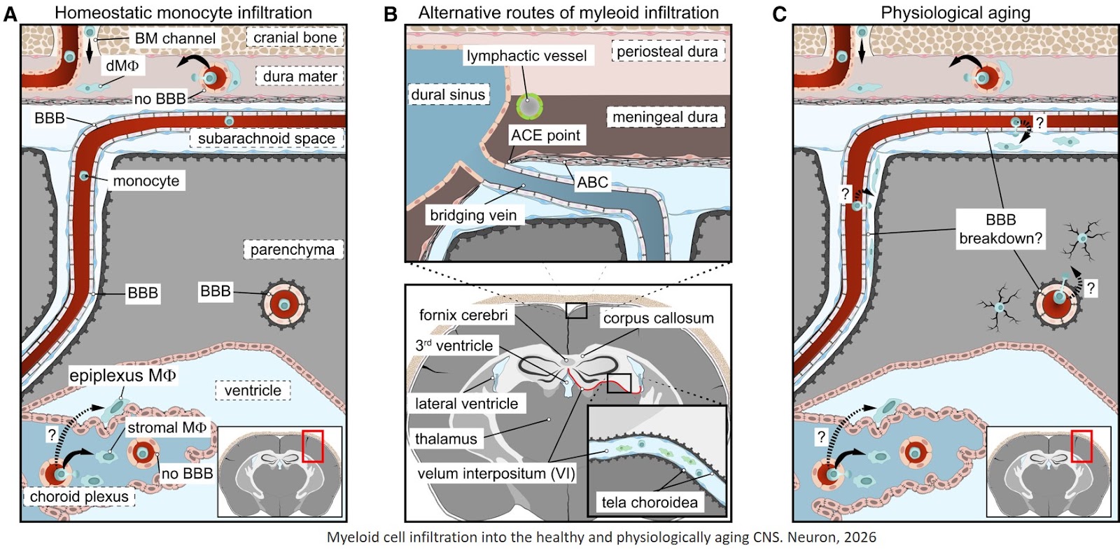
The central nervous system (CNS) regulates immune cell infiltration through highly specialized structural checkpoints known as "myeloid gateways." While the healthy, adult CNS parenchyma is typically shielded from circulating peripheral immune cells, specific developmental windows, aging processes, and neurodegenerative pathologies activate these distinct anatomical routes to facilitate entry. Understanding these gateways is crucial for designing targeted immunotherapies and mitigating chronic neuroinflammation.
During early development, long-lived resident microglia seed the brain before the blood-brain barrier (BBB) fully seals. They originate from the embryonic yolk sac and rely on specific anatomical paths to migrate into the expanding neuroepithelium:
The Pial Interface: Precursor cells (IBA1+CD206+) gather in the extraluminal cephalic mesenchyme. Mediated by the adapter protein Talin-1, these cells bind to the extracellular matrix and transmigrate directly through the pial surface into the cerebral wall.
The Ventricular/Ependymal Route: Macrophage progenitors also cross the epithelial layer of the roof plate to enter the lateral ventricles. From this cerebrospinal fluid (CSF) reservoir, they cross the ependymal layer to integrate into the developing cortex.
In adulthood, the brain restricts peripheral myeloid infiltration to maintain an optimal network environment. However, physiological stress, injury, or microglial depletion triggers recruitment through specialized border structures:
The Velum Interpositum (VI): The VI is a double-layered, vascularized fold of the pia mater tucked beneath the hippocampus. Acting as a primary "back door" to the brain, myeloid cells rapidly flood through this portal to repopulate the parenchyma if resident microglia are acutely destroyed, or during active neuroinflammatory conditions like Multiple Sclerosis (MS).
Skull Bone Marrow (SBM) Channels: Direct microscopic vascular tunnels physically connect the bone marrow of the cranial bones directly to the dura mater. Under neurodegenerative stress, bone marrow-derived monocytes and neutrophils use these internal shortcuts to bypass systemic circulation, traveling straight from the skull bone into the meninges.
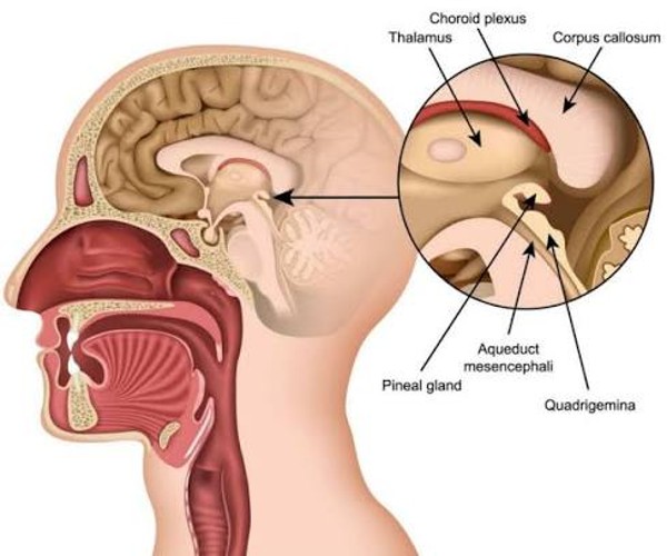
The Choroid Plexus (CP): Functioning as the blood-CSF barrier, the fenestrated capillaries of the choroid plexus serve as a prominent gateway. While its stromal macrophages are continuously replenished by bone marrow sources, systemic inflammation alters the adhesion molecules on the CP epithelium, transforming it into an active transit zone for circulating monocytes.
Rather than entering the deep brain parenchyma, certain short-lived circulating myeloid cells are selectively recruited to steady-state border zones. These cells are termed CNS-associated macrophages (CAMs) or border-associated macrophages (BAMs). They patrol external boundaries and process peripheral waste across three primary zones:
Meningeal Interfaces: Positioned within the dura, arachnoid, and pia mater layers. While dural macrophages are routinely replaced by adult bone marrow cells, deeper leptomeningeal macrophages maintain embryonic self-renewal loops.
Perivascular (Virchow-Robin) Spaces: Surrounding the major penetrating blood vessels of the brain. These cells monitor local fluid exchange along the glymphatic pathway, and their disruption often precedes full BBB leakage.