


A skin biopsy to detect a rare neurodegenerative disease
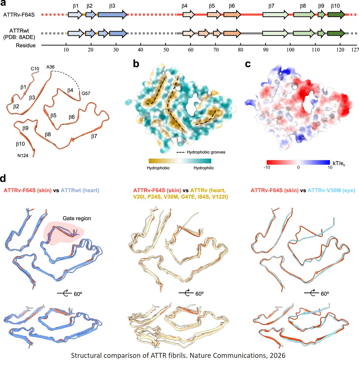
Transthyretin amyloidosis (ATTR) is a rare, progressive and highly aggressive degenerative disease. It is caused by the misfolding of a specific protein, leading to its toxic accumulation in the form of filamentous deposits in various organs.
Researchers have succeeded for the first time in determining the three-dimensional structure of abnormal protein deposits from a skin biopsy of a living patient. This breakthrough opens the door to earlier, less invasive diagnosis, potentially accessible to a much larger number of patients and applicable to other diseases. The study is published in Nature Communications.
ATTR amyloidosis is a rare, acquired or hereditary genetic disease, characterized by involvement of the nervous system, kidneys, eyes, and heart. It is caused by the misfolding of the transthyretin protein (TTR), which assembles into toxic filaments (amyloid deposits) in these organs, leading to progressive organ dysfunction. This mechanism is similar to that observed in other major neurodegenerative diseases such as Alzheimer’s, Parkinson’s, or amyotrophic lateral sclerosis.
Until now, structural studies of these fibrils were mainly based on tissues obtained after the patient’s death, consequently reflecting the terminal stages of the disease. The researchers investigated the structure of these deposits from a skin biopsy, performed quickly and almost painlessly on an ATTR patient.
Despite the small quantities, the patient’s skin sample allowed the isolation of fibrils in sufficient quantity to characterize their molecular composition and to resolve their three-dimensional structure using cryo-electron microscopy – a cutting-edge technique that enables proteins to be observed in their native state with near-atomic resolution. The researchers show that fibrils obtained from the skin biopsy have an almost identical fold to that observed in other tissues, confirming that amyloid fibrils from skin tissue faithfully reflect deposits present in organs that are more difficult to access, such as heart or brain tissues.
“The minimally invasive nature of the skin biopsy also opens new avenues for studying the disease directly in patients. It is now possible to observe how fibrils evolve over time, at different clinical stages, or in response to treatments that may alter the course of the disease,” explains a co-first author of the study. This approach makes it possible, for the first time, to consider long-term monitoring of amyloid filament structures, which could transform the way currently existing or emerging therapies are evaluated.
Encouraged by these results, the laboratory plans to apply this methodology to neurodegenerative diseases, including Alzheimer’s and Parkinson’s, which are also characterized by abnormal protein accumulation in the form of amyloid deposits.
“Being able to study the structure of deposits directly in living patients profoundly changes our ability to understand these diseases and assess the effect of treatments. It greatly expands the number of structural studies previously reserved for post-mortem samples, and thus potentially enables personalized care in the future,” concludes the author.