







How non-enveloped viruses infect cells
Viruses come in two major flavors —those enveloped in membranes, such as herpesvirus, influenza virus, HIV, and SARS-CoV-2, and those that are not, such as poliovirus, HPV, rotavirus, and adenovirus.
Scientists know more about how enveloped viruses get their genetic material across the cell membrane and into our cells, thereby causing infection, than about how non-enveloped viruses do.
Thanks to advanced imaging methods, his lab and collaborators have now filled in one of the final pieces of the puzzle for non-enveloped viruses that deliver their genomes packaged within a protein shell.
The findings, supported in part by federal funding and reported in Science, deepen understanding of virology and could help the biotechnology industry devise better methods to deliver gene therapies and other relatively large therapeutic agents into cells.
“This work is relevant not just for understanding infection by non-enveloped viruses but for addressing the general question of what are the mechanisms for delivering large cargo into cells,” said a senior author of the study. “I’ve always thought if we can figure out how evolution did it for one virus, it puts us a step ahead.”
Previous studies from the Lab and others showed how rotaviruses attach to the cell membrane and then become engulfed and brought into a membrane-bound compartment inside the cell. The virus then must somehow punch a hole in that membrane to get its DNA or RNA into the cell, take over the cell's molecular machinery, and replicate itself.
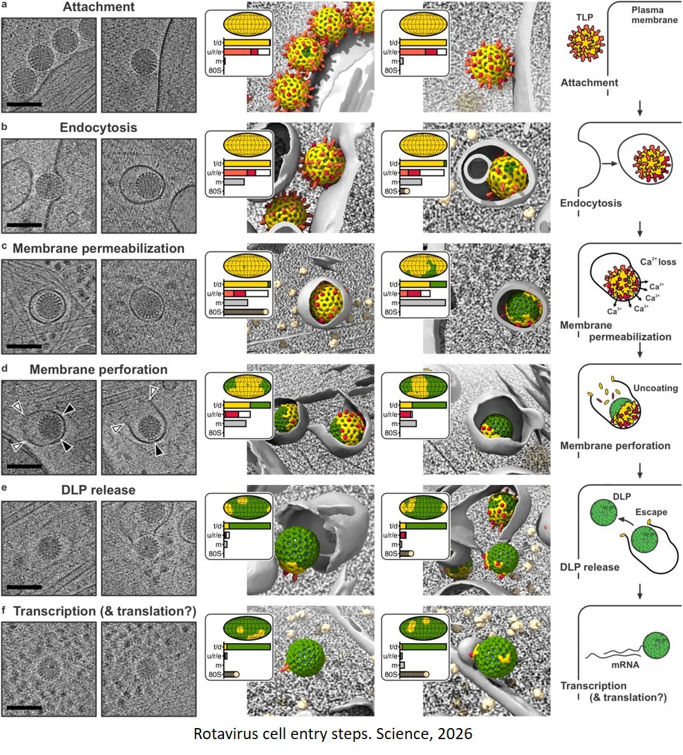
In the new study, which used a strain of rotavirus, the authors identified the protein that punches the hole in the membrane and revealed how it does so. They showed that virus protein 5, or VP5, makes the membrane permeable to calcium, causing a loss of calcium ions. This in turn makes virus protein 7, or VP7, dissociate from the virus particle and form a pore in the membrane.
“All along I had thought it would be VP5 that punched the hole, but it’s VP7,” the author said.
The team achieved this using a relatively new technology called cryo-electron tomography, or cryo-ET.
A type of cryo-electron microscopy (cryo-EM), cryo-ET allows scientists to flash-freeze an ultrathin biological sample and take high-resolution snapshots at multiple angles that can be reconstructed into a 3D model. It’s like a CT scan, only instead of the device rotating 180 degrees around a patient to capture an image, in cryo-ET the microscope stays still while the sample tilts.
The cryo-ET reconstructions delivered visuals of each step of rotavirus infection, from attaching to the cell membrane and being engulfed to punching the hole and ejecting its RNA.
An important set of experiments remains to confirm the specifics of how VP7 works, the author said.
Now that the researchers have shown how rotaviruses deliver their genetic material into cells, they plan to study the corresponding mechanisms for certain other non-enveloped viruses.