



Atomic force microscopy in bacteriology
Atomic force microscopy has enabled the high-resolution visualization of the peptidoglycan architecture in Gram positive bacteria, along with changes in this structure following antibiotics treatment.
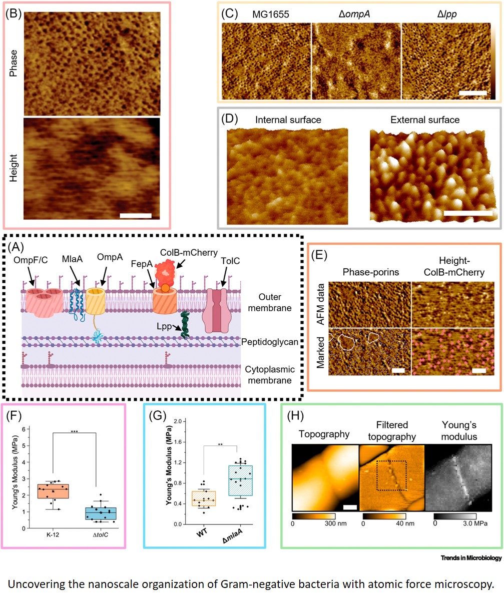
It has also led to the discovery that the outer membrane of Gram-negative bacteria is phase-separated with distinct domains rich in lipopolysaccharide or outer membrane proteins, and showed how mechanical stability of the cell envelope depends on molecular components.
Atomic force microscopy has revealed that bacterial killing by antibiotics can depend on metabolic activity and how it correlates with nanoscale changes at the bacterial surface.
Force spectroscopy accurately quantified the binding strength between adhesins and host proteins at the single-molecule and single-cell levels.
https://www.cell.com/trends/microbiology/fulltext/S0966-842X(26)00221-0