


How the brain compensates for sleep deprivation?
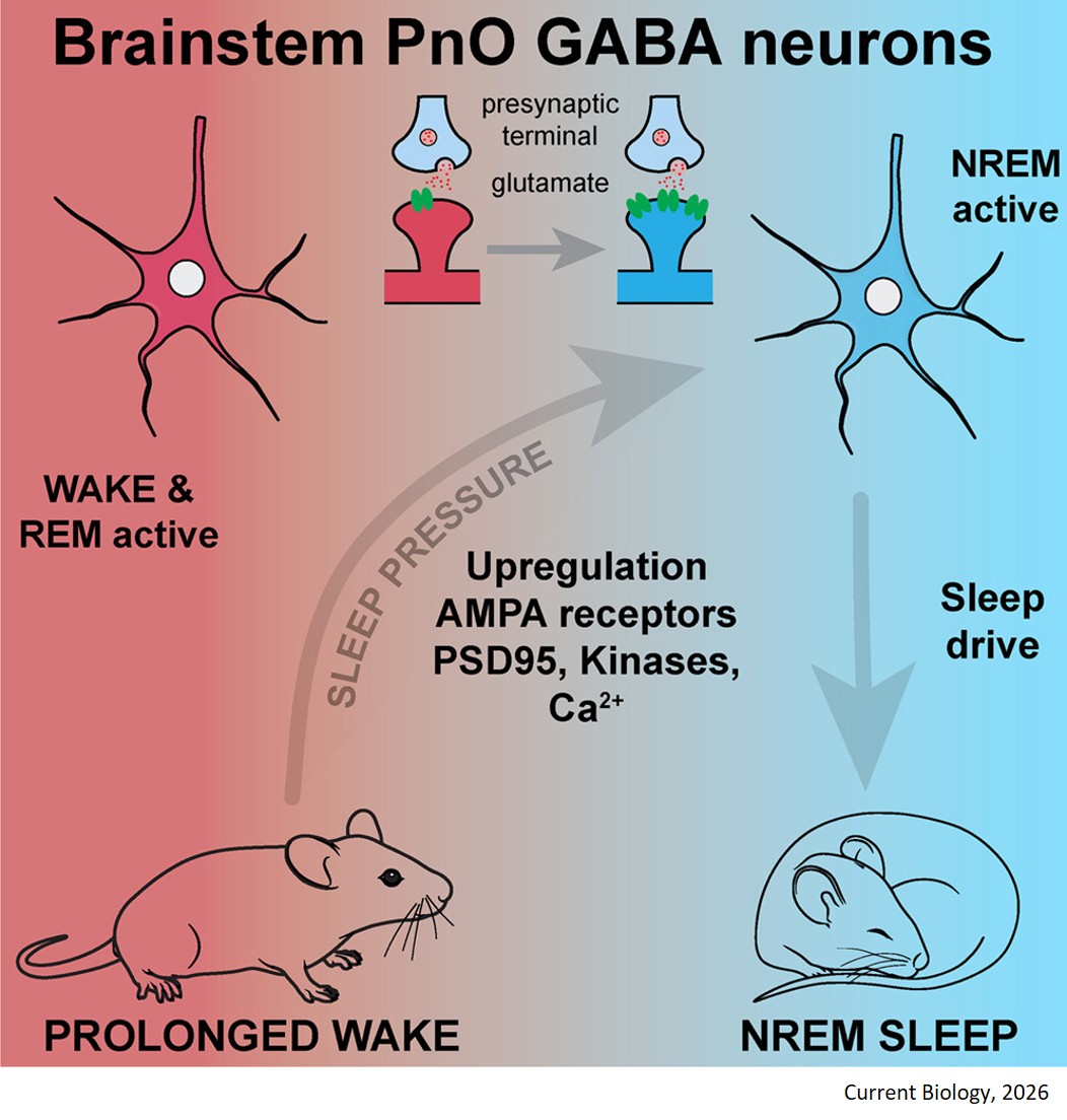
The study identified a unique population of somatostatin/parvalbumin-expressing GABAergic neurons located in the oral pontine reticular nucleus (PnOVgat). Under standard conditions, these cells are exclusively active during WAKE and Rapid Eye Movement Sleep (REMS).
As sleep debt builds, PnOVgat cells selectively accumulate GluA1-containing AMPA receptor subunits. This accumulation is paired with an increase in synaptic scaffolding proteins (PSD95) and activated protein kinases—specifically pCaMKII (T286) and pPKA (T197). These enzymes phosphorylate the critical serine residues on the GluA1 subunits, drastically increasing their excitability. The researchers confirmed a significant spike in the postsynaptic AMPA/NMDA receptor ratio, meaning the cells become hypersensitive to excitatory glutamatergic signaling accumulated during wakefulness.
Once overloaded with AMPA drive, the PnOVgat neurons adapt their firing architecture. During the first critical hour of recovery sleep, they transiently switch their activity to fire aggressively during non-REMS (NREMS).
Thus, this study demonstrates that a specific population of wake-active brainstem GABA neurons tracks sleep deprivation by upregulating AMPA receptors, acting as a homeostatic "pressure valve" to force recovery sleep (RS). This discovery provides a direct cellular mechanism for the Synaptic Homeostasis Hypothesis (SHY), proving that the brain uses localized, structural synaptic plasticity within the brainstem to monitor time spent awake and adjust subsequent sleep depth.
Uncovering how PnOVgat cells convert toxic periods of prolonged wakefulness into an automated physiological drive offers an ideal pharmacological blueprint for treating sleep fra fragmentation, trauma-induced sleep deprivation, and chronic insomnia.
https://www.cell.com/current-biology/fulltext/S0960-9822(26)00529-4