

Gene defects linked to primary ciliary dyskinesia
Hair-like appendages called motile cilia beat in synchrony on cells in many parts of the body to keep fluids and particles moving along, such as clearing mucus in the lungs so it can be coughed out.
Genetic mutations can compromise ciliary structure and function and cause disease. For instance, changes in any of more than 50 genes are known to cause primary ciliary dyskinesia (PCD), a rare disease that can damage the respiratory tract, inner ear, heart, reproductive tract, and other organs. Currently, treatments can only slow progression of PCD, not stop or cure it, and an estimated 20 to 30 percent of cases remain genetically unexplained.
A team led by researchers has now unveiled the detailed structure and composition of an understudied part of the cilium known as the transition zone — revealing that abnormalities in this zone can play a role in PCD and implicating two new genes in the disease.
The findings, published in Science, provide an explanation for at least some of the patients whose PCD has been a genetic mystery and could improve doctors’ ability to diagnose the disease. The work also offers additional genes and proteins for researchers to target as they try to develop treatments.
On a basic-science level, the study deepens understanding of how cilia work and calls scientists’ attention to the transition zone in diseases involving motile cilia.
“This is an example of how combining in situ structural biology with genetics can address fundamental questions and illuminate the causes of human disease,” said a co-senior author.
People with PCD typically have trouble clearing mucus from their lungs and sinuses and fluid from the inner ear, often leading to repeated infection, scarring, and chronic respiratory illness. Malfunctioning cilia can also cause fertility and pregnancy problems. In about half of PCD cases, one or more organs end up in the wrong places or fail to form completely during embryonic development.
Most people need to inherit two copies of a disease gene, one from each parent, to develop PCD. The disease is usually diagnosed in childhood.
The researchers study how cilia work normally and how dysfunction leads to diseases known as ciliopathies.
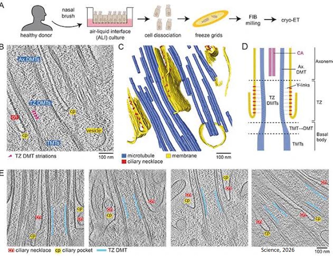
The researchers sought to pry open the secrets of the transition zone, located at the base of cilia. Mutations in this zone had been linked to diseases involving non-motile cilia, but until the new study, the mutations associated with PCD involved a different part of the cilium.
The researchers applied a cutting-edge imaging technology called cryo-FIB-ET to study cell cultures from the lining of the respiratory tract. Combined with AI tools, this allowed the researchers to capture the structure of the transition zone down to the sub-nanometer level and identify nine types of proteins.
They found that four of these proteins form complexes that link together the major components of cilia, long tubes known as doublet microtubules, in the transition zone. They then discovered that mutations in the genes for two of those proteins — ECT2L and DZANK1 — disrupt the linker complexes and lead to PCD.
The team showed that mutation-driven reductions in ECT2L and DZANK1 can change the shape of cilia — such as creating bulbous tips — and hamper their function, preventing them from beating in synchrony and clearing mucus.
The researchers suspect that this happens because the transition zone serves as a gate into the main body of cilia and the ECT2L and DZANK1 mutations cause the wrong proteins to be let in or kept out.